
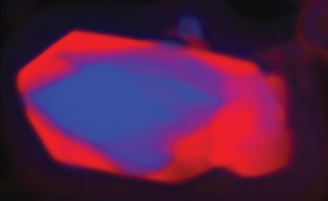
Ptychographic image using soft X-rays of lithium iron phosphate nanocrystal after partial delithiation. The delithiated region is shown in red.
A record-setting X-ray microscopy experiment may have ushered in a new era for nanoscale imaging. Working at the U.S. Department of Energy (DOE)’s Lawrence Berkeley National Laboratory (Berkeley Lab), a collaboration of researchers used low energy or “soft” X-rays to image structures only five nanometers in size. This resolution, obtained at Berkeley Lab’s Advanced Light Source (ALS), a DOE Office of Science User Facility, is the highest ever achieved with X-ray microscopy.
Using ptychography, a coherent diffractive imaging technique based on high-performance scanning transmission X-ray microscopy (STXM), the collaboration was able to map the chemical composition of lithium iron phosphate nanocrystals after partial delithiation. The results yielded important new insights into a material of high interest for electrochemical energy storage.
“We have developed diffractive imaging methods capable of achieving a spatial resolution that cannot be matched by conventional imaging schemes,” says David Shapiro, a physicist with the ALS. “We are now entering a stage in which our X-ray microscopes are no longer limited by our optics and we can image at nearly the wavelength of our X-ray light.”
Shapiro is the lead and corresponding author of a paper reporting this research in Nature Photonics. The paper is titled “Chemical composition mapping with nanometer resolution by soft X-ray microscopy.” (See below for a full list of co-authors and their affiliations.)
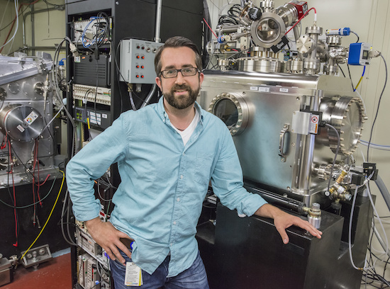
David Shapiro with the STXM instrument at ALS beamline 5.3.2.1. (Photo by Roy Kaltschmidt)
In ptychography (pronounced tie-cog-raphee), acombination of multiple coherent diffraction measurements is used to obtain 2D or 3D maps of micron-sized objects with high resolution and sensitivity. Because of the sensitivity of soft x-rays to electronic states, ptychography can be used to image chemical phase transformations and the mechanical consequences of those transformations that a material undergoes.
“Until this work, however, the spatial resolution of ptychographic microscopes did not surpass that of the best conventional systems using X-ray zone plate lenses,” says Howard Padmore, leader of the Experimental Systems Group at the ALS and a co-author of the Nature Photonics paper. “The problem stemmed from the fact that ptychography was primarily developed on hard X-ray sources using simple pinhole optics for illumination. This resulted in a low scattering cross-section and low coherent intensity at the sample, which meant that exposure times had to be extremely long, and that mechanical and illumination stabilities were not good enough for high resolution.”
Key to the success of Shapiro, and his collaborators were the use of soft X-rays which have wavelengths ranging between 1 to 10 nanometers, and a special algorithm that eliminated the effect of all incoherent background signals. Ptychography measurements were recorded with the STXM instruments at ALS beamline 11.0.2, which uses an undulator x-ray source, and ALS beamline 5.3.2.1, which uses a bending magnet source. A coherent soft X-ray beam would be focused onto a sample and scanned in 40 nanometer increments. Diffraction data would then be recorded by an X-ray CCD (charge-coupled device) and fed through a high performance data pipeline that would handle the reconstruction of the sample to a very high spatial resolution.
“Throughout the ptychography scans, we maintained the sample and focusing optic in relative alignment using an interferometric feedback system with a precision comparable to the wavelength of the X-ray illumination,” Shapiro says.
Previously, image reconstruction would have been handled by a local workstation but the very high data rates from the CCDs at the ALS make this approach no longer feasible. The high-performance data pipeline consists of a 43,000 GPU-core Infiniband Linux cluster and a dedicated high speed 10 gigabit network. It was implemented by the High Performance Computing Services (HPCS) group in Berkeley Lab’s Information Technology Division.
“This cluster executes state of art algorithms developed by the Center for Applied Mathematics for Energy Research Applications and achieves reconstruction times of only a few seconds per image,” says Gary Jung, manager of the HPCS group.
Lithium iron phosphate is widely studied for its use as a cathode material in rechargeable lithium-ion batteries. In using their ptychography technique to map the chemical composition of lithium iron phosphate crystals, Shapiro and his collaborators found a strong correlation between structural defects and chemical phase propagation.
“Surface cracking in these crystals was expected,” Shapiro says, “but there is no other means of visualizing the correlation of those cracks with chemical composition at these scales. The ability to visualize the coupling of the kinetics of a phase transformation with the mechanical consequences is critical to designing materials with ultimate durability.”
Shapiro and his colleagues have already begun applying their ptychography technique to the study of catalytic and magnetic films, magnetotactic bacteria, polymer blends and green cements.
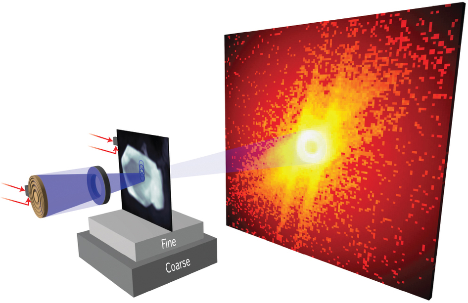
In this soft X-ray ptychography set-up, a 60 nm width outer-zone-plate focuses a coherent soft X-ray beam onto the sample, which is scanned in 40 nm increments to ensure overlap of the probed areas.
For the chemical mapping of lithium iron phosphate they used the STXM instrument at ALS beamline 5.3.2.1 which required up to 800 milliseconds of exposure to the X-ray beam for each scan. Next year, they anticipate using a new ALS beamline called COSMIC (COherent Scattering and MICroscopy), which will feature a high brightness undulator x-ray source coupled to new high-frame-rate CCD sensors that will cut beam exposure times to only a few milliseconds and provide spatial resolution at the wavelength of the radiation.
“If visible light microscopes could only achieve a resolution that was 50 times the wavelength of visible light, we would not be able to see most single celled organisms,” Shapiro says. “Where would the life sciences be with such a limitation? We are now approaching the point where we will have X-ray microscopes of comparable quality to today’s visible light instruments for the study of nanomaterials.”
Co-authoring the Nature Photonics paper in addition to Shapiro and Padmore were Young-Sang Yu, Tolek Tyliszczak, Jordi Cabana, Rich Celestre, Weilun Chao, David Kilcoyne, Stefano Marchesini, Tony Warwick and Lee Yang of Berkeley Lab; Konstantin Kaznatcheev of Brookhaven National Laboratory; Shirley Meng of the University of San Diego; and Filipe Maia of Uppsala University in Sweden.
This research was primarily supported by the DOE Office of Science.
Additional Information
For more about the Advanced Light Source go here
For more about David Shapiro’s research go here
# # #
Lawrence Berkeley National Laboratory addresses the world’s most urgent scientific challenges by advancing sustainable energy, protecting human health, creating new materials, and revealing the origin and fate of the universe. Founded in 1931, Berkeley Lab’s scientific expertise has been recognized with 13 Nobel prizes. The University of California manages Berkeley Lab for the U.S. Department of Energy’s Office of Science. For more, visit www.lbl.gov.
DOE’s Office of Science is the single largest supporter of basic research in the physical sciences in the United States, and is working to address some of the most pressing challenges of our time. For more information, please visit the Office of Science website at science.energy.gov/.