
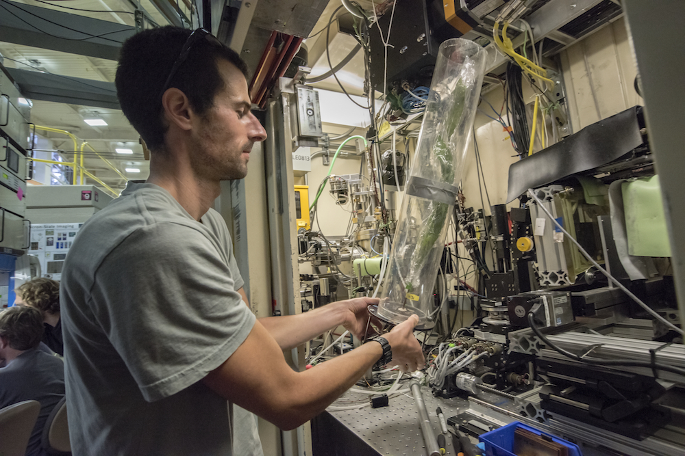
Thorsten Knipfer, a UC Davis postdoctoral researcher, carries an American chestnut sapling into the experimental chamber at Berkeley Lab’s Advanced Light Source. Knipfer and other researchers used X-rays at ALS Beamline 8.3.2 to scan the stems of dozens of saplings. The work will produce 3-D reconstructions of the stems’ microscopic structure. (Credit: Marilyn Chung/Berkeley Lab)
To resolve open questions about plant plumbing—how plants transport water from roots through stems and how they respond to stress such as drought—science teams from around the world met Sept. 1-3 at the Department of Energy’s Lawrence Berkeley National Laboratory (Berkeley Lab) and UC Berkeley for an intensive round of experiments using X-rays and other techniques.
The collaborative effort to study plants prepared under different watering conditions will provide new data and insight about how to better tend to crops and other plants under stress and in natural conditions. It could also lead to improved understanding and forecasting for drought-related die-offs of trees and other plant species.
About 15 researchers participated in the event, including scientists from the University of California, Davis; Western Sydney University in Australia; and the University of Alberta in Canada; and together they will work to analyze the data and interpret the results.
Seeing inside plant stems in 3-D
Different study methods have so far yielded different results about how plants move water and repair blockages in water flow.
Researchers studied live American chestnut saplings with a 3-D microscopic X-ray imaging technique at Berkeley Lab’s Advanced Light Source (ALS), and then explored the same samples using other laboratory tests.
“For me this is unprecedented—something totally different,” said Dula Parkinson, the ALS scientist who oversees the X-ray beamline and facilitated the effort to scan the lower stems of dozens of the young trees with X-rays. “We don’t usually have this many people from this many groups coming here all at the same time. It’s really been fun.”
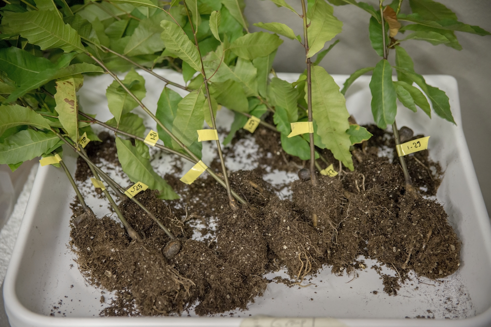
These American chestnut saplings, grown at a nursery in Idaho, are prepared for a round of X-ray and other experiments at Berkeley Lab’s Advanced Light Source. (Credit: Marilyn Chung/Berkeley Lab)
Typically, just a handful of researchers at a time participate in an experiment at a single ALS experimental station. The ALS has developed an expertise in scanning different parts of plants, including stems, leaves, roots, berries and flowers, using a technique known as microtomography, which produces 3-D renderings at microscopic resolution.
The chestnut trees were grown and prepared for the experiments at a University of Idaho nursery. The samples were separated into different groups based on how much water they received prior to the experiments. Each tree was wrapped in plastic to preserve its moisture during each X-ray scan, which lasted about 12 minutes as each tree was rotated 180 degrees in the focused X-ray beam.
Understanding plants’ response to drought conditions
Andrew McElrone, a research scientist with the U.S. Department of Agriculture’s Agricultural Research Service and an associate adjunct professor at UC Davis, said that drought conditions are a source of stress that can create gas bubbles in plants’ water transport systems, which can lead to blockages.
Scientists want to understand how plants repair themselves or are permanently damaged by this process, so they have used X-rays to study the internal structure of plants’ stems. Other studies included the forcing of water through cut plant stems to observe how efficiently they transport water in response to varying conditions.
“The formation of bubbles under stress leads to hydraulic dysfunction,” McElrone said. Similar to our own circulatory system, if there are severe blockages in their vessels then the plants die. Some plants are better than others at repairing their vessels and surviving these blockages.
McElrone has led earlier ALS studies of water transport in grapevines that have stirred discussions in the research community about the utility of X-rays for studying plant hydraulics.
Brendan Choat, a senior research lecturer at Western Sydney University in Australia who participated in the working group, said, “The work we are doing here will give us a much clearer picture of how we should be measuring drought tolerance in plants and which measurements should be included in mortality models.”
The results could help improve modeling and forecasting for tree mortality, for example, he said, adding, “Mechanisms of plant mortality are all interdependent: water stress is a primary factor and this can lead to increased susceptibility to pests and pathogens.”
Multiple methods seek out root of problems in plant damage, die-offs
One question researchers hoped to resolve in this latest round of X-ray and conventional water-transport studies is whether the X-rays themselves, or sample preparation from more conventional laboratory techniques, impact the measurements in any way. The studies could show whether X-ray studies can potentially undercount blocked vessels, and also whether hydraulics studies using cut stems can introduce additional air bubbles or otherwise alter natural samples and give false readings.
“We’re trying to come up with the best way to combine and interpret different sets of data,” said Anna Jacobsen, associate professor of biology at California State University, Bakersfield, who participated in the working group.
Jacobsen examined stem cross-sections with a microscope in an ALS conference room that was converted into a makeshift plant laboratory for the research event. The samples were fed blue-dyed water to easily identify which vessels were transporting water, which is key in moving sap through healthy plants, and those that appeared blocked by bubbles or other types of obstructions.
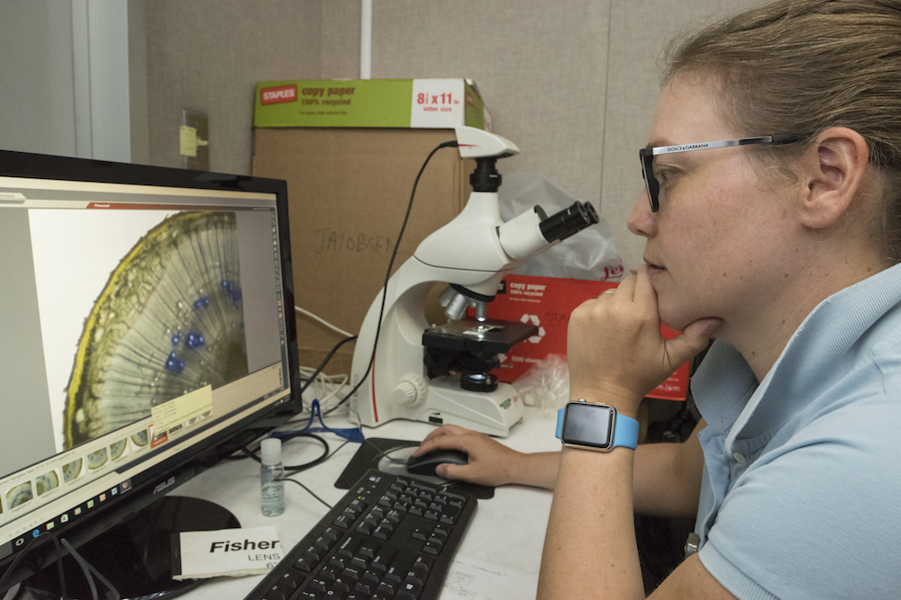
Anna Jacobsen, associate professor of biology at CSU Bakersfield, studies water uptake in cut stems during a research event hosted by Berkeley Lab’s Advanced Light Source. (Credit: Marilyn Chung/Berkeley Lab)
For Jacobsen and colleague Brandon Pratt, a professor of biology at California State University, Bakersfield, who specializes in studies of Southern California shrubland species that are particularly impacted by drought, the working group was their first experience working at an X-ray source.
“This will provide proof about some of the methods we’re applying,” Pratt said. “We are using three independent methods to measure the samples in different ways.”
Martin Venturas, a postdoctoral researcher at the University of Utah, worked at a separate station to measure cut samples in a lab setup known as a conductivity apparatus that measured the flow of water through each stem segment.
The technique, which factors in the length and width of each segment, can inform researchers about the maximum flow of liquid through a given stem section if all of the section’s vessels are active in water transport. It can also detect when flow declines due to gas bubbles.
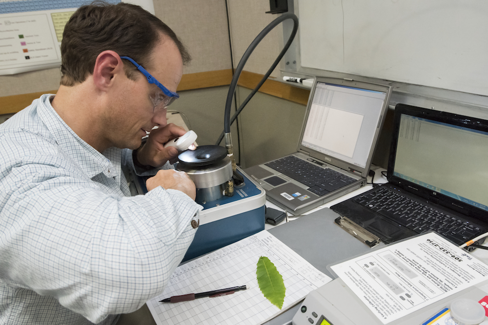
Brandon Pratt of CSU Bakersfield examines a stem segment at Berkeley Lab’s Advanced Light Source. (Credit: Marilyn Chung/Berkeley Lab)
After the X-ray and benchtop studies at the ALS, the samples were transported to the UC Berkeley campus for study in a centrifuge, a rapidly rotating machine that can simulate the stress that accompanies drought conditions.
McElrone noted that the working group follows a National Science Foundation workshop held last year that also united researchers around common research questions in plant hydraulics. The latest event produced a terabyte worth of 3-D imaging data that will be processed and stored at Berkeley Lab’s National Energy Research Scientific Computing Center (NERSC) and ultimately shared with the greater research community.
The ALS and NERSC are DOE Office of Science User Facilities.
The organizing committee for the event included: Andrew McElrone and Thorsten Knipfer of UC Davis, Brendan Choat of University of Western Sydney, Craig Brodersen of Yale University, Daniel Johnson and Z. Carter Berry of University of Idaho, and Duncan Smith of University of Wisconsin, Madison.
Researchers from UC Davis; California State University, Bakersfield; University of Idaho; Yale University; University of Wisconsin; University of Utah; University of West Sydney in Australia; and University of Alberta in Canada were among the participants in the ALS event. This work is supported by the National Science Foundation and the Department of Energy’s Office of Basic Energy Science.
###
Lawrence Berkeley National Laboratory addresses the world’s most urgent scientific challenges by advancing sustainable energy, protecting human health, creating new materials, and revealing the origin and fate of the universe. Founded in 1931, Berkeley Lab’s scientific expertise has been recognized with 13 Nobel prizes. The University of California manages Berkeley Lab for the U.S. Department of Energy’s Office of Science. For more, visit www.lbl.gov.
DOE’s Office of Science is the single largest supporter of basic research in the physical sciences in the United States, and is working to address some of the most pressing challenges of our time. For more information, please visit science.energy.gov.