Berkeley Lab at ACS Fall Meeting 2010: Environment
As evident by the proliferation of weekend garage sales and flea-markets, it seems almost human nature to “junk up” our domicile until the point is reached where we have to remediate. Our planet is no exception as we have polluted land, air and sea through our activities to the point where environmental remediation is a major enterprise. Among the Berkeley Lab presentations at the ACS Fall 2010 meeting were two reports describing unique technological approaches to this enterprise.
Medical Imaging for the Environment
 In a talk titled “Applications of PET imaging toward bioremediation and geochemical studies,” Nicholas Vandehey, a medical physicist with Berkeley Lab’s Life Sciences Division’s Biomedical Isotope Facility, discussed the application to bioremediation and carbon sequestration studies of imaging technologies usually associated with medicine – positron emission tomography (PET) and single photon emission computed tomography (SPECT).
“In bioremediation research, which has a focus on immobilizing or eliminating contaminants from soil, many experiments are done in large columns through which ground water mixtures are passed and data is collected by taking discrete samples on ports along the length of the column,” Vandehey said. “We’re looking to improve on these methods by presenting the capability of dynamic imaging of the whole volume of the column, an idea that is otherwise unheard of in bioremediation but routine to nuclear medicine.”
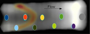
Vandehey described one study of sediment from a site near the town of Rifle, Colorado, where the soil is contaminated with uranium from nearby mills. A bioremediation plan has been proposed whereby bacteria would be used to feed on the contaminants and degrade them to an immobile form. Vandehey and his collaborators used technetium 99m, a single photon-emitter, to label pertechnetate, which was then passed through soil samples. They used SPECT imaging to monitor the reduction of the pertechneate by iron(II) from a state that is highly mobile in water to an immobilized state. Iron(II) is a byproduct of the microbe’s metabolism.
“We injected the technetium into three vials, the first filled with ground water, the second with ground water and an acetate-augmented rifle sediment, and the third with non-augmented sediment,” Vandehey said. “We ran five serial studies, shaking the vial between each study, with increasing time between shakes. Regions of interest were drawn to encompass the non-sediment portion of each vial so as to quantify the rate of removal of the technetium from the solution.”
With SPECT imaging, Vandehey and his colleagues were able to successfully use the technetium to track the rate of immobilization of pertechnetate, which was only seen in the sample with active bacteria.
“Sampling ports in traditional bioremediation studies allow for monitoring of column conditions, but can only probe the column at select locations at discrete time points,” Vandehey said. “Our approach provides dynamic measurements of the entire column volume.”
Vandehey said this approach could be valuable for remediation projects such as the decontamination of the Hanford site in Washington, or for getting a better understanding of the role of redox chemistry in environmental remediation.
In his talk Vandehey also described a study in which carbon-11, a radioactive form of carbon with a relatively short half-life that makes it particularly useful in PET imaging, was used to take a new look at carbon sequestration. The idea of capturing carbon dioxide at the point of emission rather than allowing it to escape into the atmosphere has gained traction as a tool for combating global climate change. However, this captured carbon must then be safely stored or sequestered. Proposals have called for storage underground or deep in the ocean, or for mineral carbonation into stable calcium and magnesium carbonates. However, each of these approaches raises concerns about the stored carbon leaking back into the atmosphere.
“To demonstrate how medical imaging can be applied to carbon sequestration, we performed an experiment passing carbon dioxide gas over ascarite to trap the gas, as would be done in carbon sequestration,” Vandehey said. “In this case, the ascarite, which is sodium hydroxide on silicate, reacts with the carbon dioxide to form water and sodium carbonate, with a capacity of about 30-percent carbon trapping by weight.”
Vandehey and his colleagues passed carbon-11 labeled carbon dioxide through an empty column and into an ascarite plug in a second column, then used PET to image the columns for 80 seconds with five second duration frames.
“The images from this experiment were exactly as expected,” Vandehey said, “with the radio-labeled gas slowly passing through the first column and immediately being trapped on the initial portion of the second column, right where the ascarite starts.”
Vandehey also described a mineral carbonation experiment just getting underway in which carbon dioxide is reacted with magnesium chloride to yield magnesium carbonate plus hydrogen chloride. The idea is to bubble the radio-labeled carbon dioxide through solutions and measure the percentage of trapped carbon as a way to quantify carbonation.
“In the hope of enticing future collaborations,we’re mainly looking to demonstrate our capabilities with radioisotope imaging methods for determining biological characteristics of bacteria, oxidation states of contaminant metals, and other aspects of environmental remediation,” Vandehey said.
Synchrotron Light and Organic Aerosols
 In another departure from traditional environmental sample studies, Christopher Cappa, a professor of environmental engineering at the University of California, Davis, and a guest researcher at Berkeley Lab’s Advanced Light Source (ALS), discussed the use of synchrotron radiation, specifically vacuum ultraviolet (VUV) light, to study how the properties of aerosols in Earth’s atmosphere vary in both space and time.
“Chemical composition plays an important role in determining the lifetime and climate impacts of atmospheric aerosols,” Cappa said. “The efficiency with which aerosols interact with light depends importantly on both particle composition and size. Our studies have focused on the evolution of laboratory-generated organic aerosol optical properties upon heterogeneous oxidation by hydroxide radicals.”
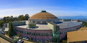
For this work, Cappa and his collaborators utilized the world’s only VUV aerosol mass spectrometer, which is stationed at ALS Beamline 9.0.2, aka the “Chemical Dynamics Beamline.” The vacuum ultraviolet light generated by an undulator magnetic device enables the Chemical Dynamics Beamline to serve a broad range of chemical physics research, with unique applications to certain specific areas such as aerosol science. Cappa and his collaborators, which included Berkeley Lab’s Kevin Wilson and Jared Smith, of the Chemical Sciences Division, used this beamline and its facilities to investigate how heterogeneous oxidation affects the optical properties of organic aerosols. They then correlated their observations with aerosol chemical compositions.
“We showed that both particle extinction and particle hygroscopicity continuously increase with increasing hydroxide exposures,” Cappa said. “We also found that differences in the observed response to oxidation are related to the starting aerosol identity, for example whether the aerosol is hydrophobic or hydrophilic. This indicates the importance of water solubility on hygroscopicity.”