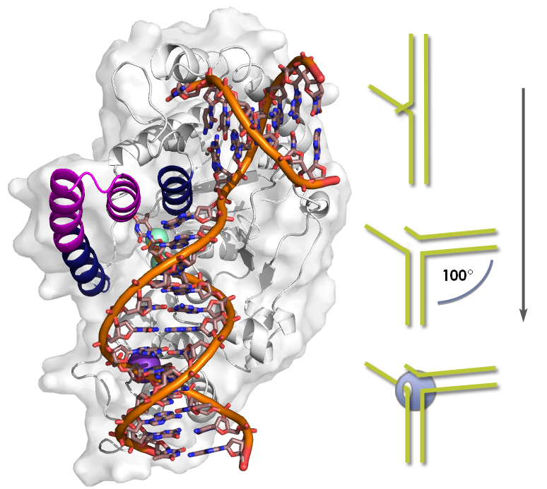
During DNA replication of the lagging strand, numerous Okazaki fragments must be joined. The newer fragment ends in a short flap call the 3’ overhang, while the previous fragment leaves a long 5’ flap after its primer is removed. The junction opens when the template strand is bent 100 degrees. FEN1 grasps the DNA at the bend, threads the flap through an archway, and trims the flap to match the overhang. (Click on image for best resolution.)
DNA replication is critical to the life of all organisms, insuring that each new cell, as well as each new offspring, gets an accurate copy of the genome. Among the legions of proteins that do the work so essential to a cell’s survival, the DNA-slicing “flap endonuclease” FEN1 plays a key role.
The structure of human FEN1 has now been solved by an international team of scientists led by the U.S. Department of Energy’s Lawrence Berkeley National Laboratory (Berkeley Lab) and the Scripps Research Institute in La Jolla (Scripps). The structure reveals the surprising mechanism behind FEN1’s speed, accuracy, and versatility.
“FEN1 has to perform 50 million operations during each replication. It has to do them quickly and it can’t be sloppy,” says John Tainer of Berkeley Lab’s Life Sciences Division (LSD) and Scripps. “But FEN1 is also important in DNA repair, which presents different challenges. Its ability to target specific repair pathways makes it of urgent interest in cancer research.”
“We wanted to know how FEN1 can do different jobs, and how the larger family of which it’s a member can perform similar functions on very dissimilar arrangements of DNA,” says Susan Tsutakawa of LSD, first author of a paper in the journal Cell describing the team’s work. “The secret of how FEN1 interacts with DNA lies in its structure.”
In the beginning, replication
When a cell reproduces itself, the critical first step is DNA replication, which starts by unwinding double-stranded DNA. The resulting single-strand “templates” are then processed by intricate cellular machinery to form two duplicates of the original double helix.
Much of the work is done by a protein called DNA polymerase, which adds nucleotides complementary to the template’s nucleotides. But polymerase can’t build the copy without help; first an RNA primer has to bind to the template with a few base pairs, acting as a short length of “pseudo-DNA” to whose backbone the polymerase can start adding DNA nucleotides to match with the template. The polymerase keeps on adding nucleotides until it runs into the RNA primer from the previous start.
This doesn’t happen often on the so-called leading strand of the unwound DNA, where the polymerase usually proceeds smoothly, as if on a one-way street. The one-way direction, named for the chemical composition of the nucleotides at opposite ends of a DNA strand, is 5’ to 3’ (five-prime to three-prime).
On the lagging strand, however, the one-way street runs in the opposite direction, so replication has to be done in many little discrete fragments assembled “backwards,” each started by an RNA primer. Called Okazaki fragments, these are only about 100 nucleotides long in humans, and some 50 million of them are added to the lagging strand during a human cell’s replication.
When polymerase runs into the RNA primer of a previous fragment it peels it away like a chisel, leaving a long tail or “flap” of excess single-strand DNA at the 5’ end. This flap must be clipped off before the new fragment can be joined – failure to do this accurately would leave a gap or overlap that could cause a genetic mutation or rearrangement resulting in damaged chromosomes. FEN1 cuts off the flap and precisely prepares it for joining to the newer fragment, which is also left with a tiny flap, known as the 3’ overhang.
Much of the FEN1 structure was solved by Sakurai et al, but how FEN1 works was not apparent in the DNA-free structure. The presence of DNA appears to induce the transition from disorder to order; FEN1 positions the 5’ flap and the 3’ overhang mainly by grasping the double-strand portions of DNA on either side of the 100-degree bend.
Just how does FEN1 do the job? Tainer notes that many important structural features of human FEN1 were known from studies in 2007 by Shigeru Sakurai of Japan’s Nara Institute of Science and Technology and from work on related FEN from microorganisms known as archaea. These studies were done without FEN1 acting on DNA, however, and many questions remained.
For example, FEN1 was long thought to move into position by sliding down the 5’ flap to the incision site, like a bead on a string. The 5’ flap passes through an opening between two helical coils of amino-acid residues in the protein, which form an arch. But whether the end of the 5’ flap threads through this arch or whether the arch closes around the flap was unknown. The Sakurai structure couldn’t answer the question because this area of FEN1 was disordered in the model, with its associated structures free to move.
Considering FEN1’s remarkable specificity and accuracy, Tainer saw that neither its structure nor mechanism could be solved unless DNA with a 5′ flap was also present. Determining the structure of the two together was essential.
Solving the compound structure
Using the SIBYLS beamline at Berkeley Lab’s Advanced Light Source (ALS), designed specifically to tackle the kind of problem the FEN1 structure presented, Tsutakawa worked with Scott Classen at the ALS and Andy Arvai at the Scripps Institute. (SIBYLS stands for Structurally Integrated Biology for Life Sciences.) They rapidly obtained some 20 structures of FEN1 bound to DNA and used PHENIX software developed by Paul Adams of Berkeley Lab’s Physical Biosciences Division to derive models of three conformations of DNA in the grip of FEN1. With the DNA in place, it was possible for the researchers to see what wasn’t visible in the Sakurai model of FEN1.
“We saw that FEN1 binds the double-strand DNA on either side of the 5’ flap and opens it by severely bending the template strand all the way back to 100 degrees, almost at right angles,” says Tsutakawa. “Only because there’s a break on one side of the double-stranded DNA at that point can the DNA bend that sharply.”
“FEN1 is shaped roughly like a left-handed boxing glove,” Tainer says, noting that FEN1 holds DNA at four binding sites, beginning with two widely separated regions where it grips the double-strand DNA by its template strand. One of these binding sites is near the tip of the glove, grasping the 3’ overhang at the break so as to expose a single unpaired nucleotide. The other double-strand binding site is at the wrist of the boxing glove, near the 5’ flap.
Two other sites hold the template strand opposite the open junction and, crucially, at the crease between thumb and forefinger, where the 5’ flap will be cut.
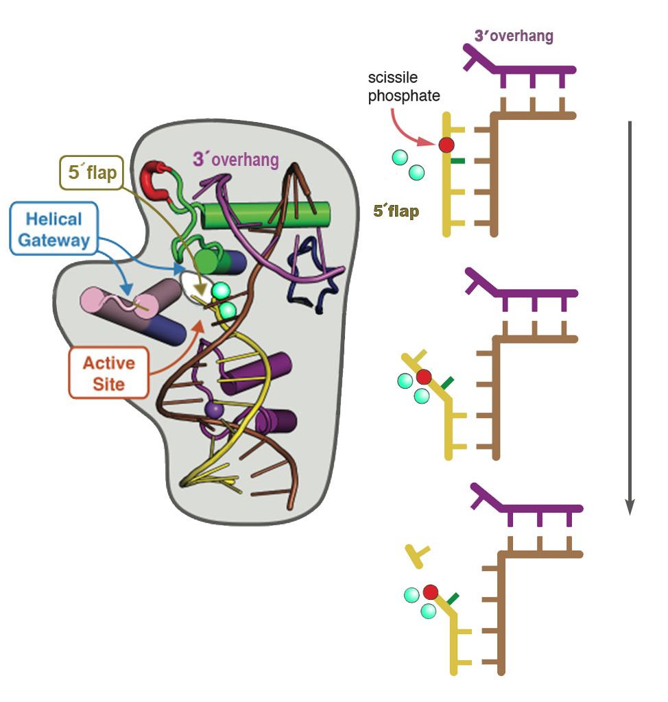
In human FEN1, the arch between the thumb and the rest of the glove is indeed capped, although this is not true for other members of FEN1’s family. In FEN1 the 5’ flap is forced to thread through the arch, which is too narrow for double-strand DNA to enter and thus selects for the single-stranded flap.

FEN1 threads the 5’ flap through the narrow archway until it is positioned so that the new strand’s backbone can be sliced between two already paired bases. Two catalytic metal ions (green spheres) stabilize and position the “scissile phosphate” for attack by a hydroxide. The fragments are now prepared for direct ligation (joining) into a continuous double strand, which replicates the original double helix. (Click on image for best resolution.)
The flap is cut at a place called the “scissile phosphate.” Phosphates (and sugars) are like the vertebrae of a single strand’s backbone, to which the bases are anchored (a phosphate plus a base makes one nucleotide). The flap is held firmly in the right place for cutting because the nucleotide with the scissile phosphate, plus its neighboring nucleotide, are initially base-paired to the template strand. The bend in the template strand opens these pairs and lines up the target nucleotides near two metal ions, which catalyze the hydrolysis – the clipping – of the flap at the precise scissile-phosphate location.
“The structure was unexpected, but it made clear how FEN1 works,” says Tsutakawa. “At the same time it explains how the superfamily of similar endonucleases can operate in such different situations.”
Motion and precision
“How does FEN1 contribute to recombination and repair accurately, without mistakes?” Tainer asks. “We see how moving the template will position the 5’ flap for precise cutting. Incision is equally precise. FEN1 cuts it where it knows the strand has already been accurately paired to the template, leaving just one unpaired base, which can then pair with the unpaired 3′ overhang.”
Tsutakawa summarizes the findings: “FEN1 grasps the template of the double-strand DNA where the junction is bent 100 degrees. The helicity of the double strand positions the 5’ flap, threading it through the arch. The helicity of the double strand, plus the binding of the 3’ overhang, also prompts the disordered parts of FEN1 to close up. Finally two paired bases on the 5’ flap are pulled apart over the metal ions, triggering incision at the scissile phosphate.”
She adds, “Many biologists still have the model of FEN1 sliding down the 5’ flap in their heads, so our model – with no sliding, but rather pushing the 5’ flap through the narrow arch – is ‘Copernican.’ It requires a whole new way of looking at this pivotal protein activity.”
Says Tainer, “Knowing how the parts move, we know how we can interfere with them. FEN1 plays a dual role in cancer – it is essential to health, but it’s also overproduced in many cancers and associated with aggressive tumors. By designing drugs that induce changes in specific structures – rather than active sites which may be nonspecific – we have hope for more effective, more targeted, and less damaging treatment.”
Additional information
“Human flap endonuclease structures, DNA double base flipping and a unified understanding of the FEN1 superfamily,” by Susan E. Tsutakawa, Scott Classen, Brian R. Chapados, Andy Arvai, L. David Finger, Grant Guenther, Christopher G. Tomlinson, Peter Thompson, Altaf H. Sarker, Binghui Shen, Priscilla K. Cooper, Jane A. Grasby, and John A. Tainer, appears in the 15 April 2011 issue of the journal Cell. This work was supported by the National Institutes of Health/National Cancer Institute’s Structural Cell Biology of DNA Repair Machines (SBDR) program project, whose co-principal investigators are John Tainer and Priscilla Cooper of Berkeley Lab’s Life Sciences Division.
The Advanced Light Source (ALS) and Stanford Synchrotron Radiation Lightsource (SSRL) are supported by the DOE Office of Science. Structural data on the FEN1 protein was collected at the ALS SIBYLS beamline (12.3.1) and at the SSRL beamline 11-1, which are supported by the National Institutes of Health/National Center for Research Resources and DOE’s Office of Science. For more information on the ALS visit http://www-als.lbl.gov/.
Lawrence Berkeley National Laboratory addresses the world’s most pressing scientific challenges by advancing sustainable energy, protecting human health, creating new materials, and revealing the origin and fate of the universe. Founded in 1931, Berkeley Lab’s scientific expertise has been recognized with 12 Nobel prizes. The University of California manages Berkeley Lab for the U.S. Department of Energy’s Office of Science. For more, visit http://www.lbl.gov/.