Scientists studying the human tissues and entire living model organisms have an array of tools at their disposal to view the inner workings of our biological systems, from mass spectrometry imaging and optical microscopies, which can make pictures of entire tissues and organs, down to X-ray crystallography and NMR (nuclear magnetic resonance), which can image single molecules. Yet no scientist is fully versed in the full scale of imaging techniques.
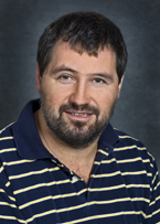
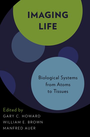
 Now Lawrence Berkeley National Laboratory scientist Manfred Auer has helped put together a book that he believes is the first to include all the biological imaging techniques up to the tissue level in one place. Imaging Life: Biological Systems from Atoms to Tissues, published by Oxford University Press, covers a wide scale of biological imaging and was edited by Auer, Gary C. Howard of the Gladstone Institute, and the late William E. Brown of Carnegie Mellon University.
“We believe imaging is the next frontier. We hope this book will help catalyze further discussion about integrated bioimaging,” Auer said. “This is the first time that one single book describes all the techniques. We can compare them to each other, their strengths and weaknesses.”
Imaging is crucial, Auer says, in order to truly understand life from a “systems biology” viewpoint. “Omics—genomics, metabolomics, proteomics—only gives you a parts list. It’s basically a phone book, with millions of names organized into tables,” he said. “What’s missing is a sort of Google maps. We’re really only going to understand how life is organized if we include the spatial relationships.”
Imaging Life is not a textbook, but rather, is meant for students and others as an introduction to the field as well as for experts in one field interested in learning about other fields, Auer said.
 “For example, people who do imaging at the single molecule level do not understand techniques focusing at the mesoscale and vice versa,” he said. “The key is for people that, like us, have an understanding in one particular area of imaging, to understand how the other fields complement what they’re doing. So it’s meant for other researchers, but also for students so they can get an idea what the other disciplines are.”
Berkeley Lab scientists who contributed to the book include Hoi-Ying Holman and Liang Chen, who co-wrote a chapter on synchotron FTIR spectral imaging; Martin Schmidt, Pradeep Parera, Alexander Weber-Bargioni, Paul D. Adams, and P. James Schuck, who co-wrote a chapter on Raman spectroscopic imaging; and Elizabeth A. Smith, Bertrand P. Cinquin, Gerry McDermott, Mark A. Le Gros, and Carolyn A. Larabell, who co-wrote a chapter on soft x-ray tomography. Auer also wrote the chapter on electron tomography and advanced 3D SEM imaging,
Berkeley Lab launched the Integrated Bioimaging Initiative several years ago as a way for researchers with expertise across the broad spectrum of imaging technologies to exchange ideas and knowledge and address the bottlenecks in integrated bioimaging, allowing them to go beyond mere pictures and actually yield quantitative information, thus ushering in a new era of “spatial systems biology.”
The book is available on Amazon.
