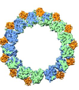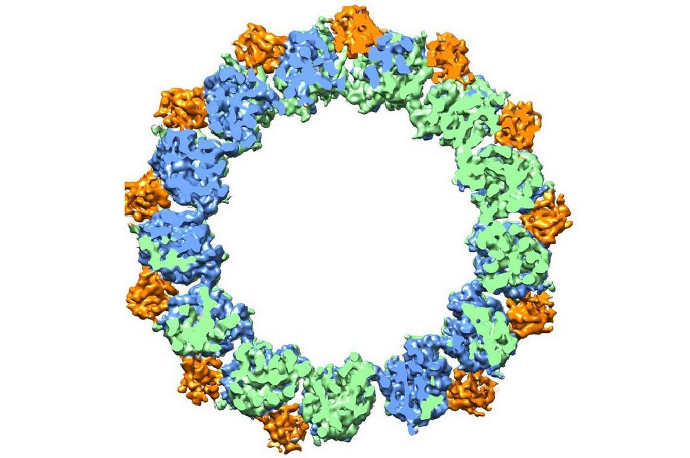
High-resolution cryoEM imaging and a unique analysis tool enabled this image of a microtubule, a hollow cylinder with walls made up of a mix of tubulin proteins. (Credit: Berkeley Lab)
A “Future Electron Microscopy” workshop held Tuesday at the ALS User Support Building showcased the breadth and depth of electron microscopy at Berkeley Lab. In addition to the recent advances in imaging a broad range of materials and biological samples at atomic, or near-atomic scales, the workshop highlighted the many opportunities that could come from integrating capabilities across the Laboratory.
The daylong workshop also chronicled the Lab’s pioneering history in pushing the state of the art in atomic resolution electron microscopy (EM), and featured a dozen presentations on science enabled by a range of electron microscopy-based techniques, such as cryoTEM (transmission electron microscopy at cryogenic temperatures), atomic-resolution tomography, in situ microscopy techniques and low energy electron microscopy.
In addition, a half dozen presentations on ultrafast electron sources, high-speed electron detectors, algorithms and supercomputing highlighted the benefits to the field of electron microscopy enabled by the diverse capabilities at Berkeley Lab.
Deputy Director Horst Simon launched the workshop with the goals of identifying gaps in technologies, finding synergies among different areas of research, and identifying future growth areas.
“What we really want to get out of this workshop is to find new ways that we can work together … and what we should be doing but that we’re not doing,” he said.
Simon noted the Lab’s significant contributions to electron microscopy through the Molecular Foundry’s National Center for Electron Microscopy (NCEM), and that several Lab scientists who have been recognized with Department of Energy Early Career Research Program awards are working with, or improving upon, electron microscopy.

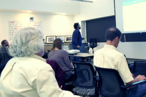
Daniele Filippetto (top and middle), a Berkeley Lab scientist, discusses his work in ultrafast electron diffraction during an electron microscopy workshop. (Credit: Berkeley Lab)
Robert Glaeser, a lab senior scientist in the Molecular Biophysics & Integrated Bioimaging Division, and a UC Berkeley professor emeritus of biochemistry, biophysics and structural biology, related the formative years of cryoEM research particularly the importance of freezing samples to liquid nitrogen temperatures to help preserve them for EM studies and stave off damage from intense electron beams, what is now known as cryoEM.
Simon also noted that the Lab’s facilities, combined with its scientific expertise and experience, have created the potential to leverage and build upon its ability to continue to make pioneering advances in this field, which is becoming increasingly important to several scientific disciplines.
The workshop was organized by Peter Denes, Paul Adams and Andy Minor. Paul Adams, Director of the Molecular Biophysics & Integrated Bioimaging Division, noted that the recent revolution in electron microscopy for biosciences has opened up many new avenues of research and exciting synergies with non-biosciences programs at the Lab.
Andy Minor, Director of the National Center for Electron Microscopy in the Molecular Foundry, noted that the most recent advances in EM moved us from 2-D to 3-D atomic-scale resolution, and that the development of new fast detectors enabled the direct mapping of properties and imaging techniques weren’t accessible before.
Minor added, “Developing the capability to provide users the best resolution at any given time scale would be a major new development for exploring dynamic processes in materials with electron microscopy.”
