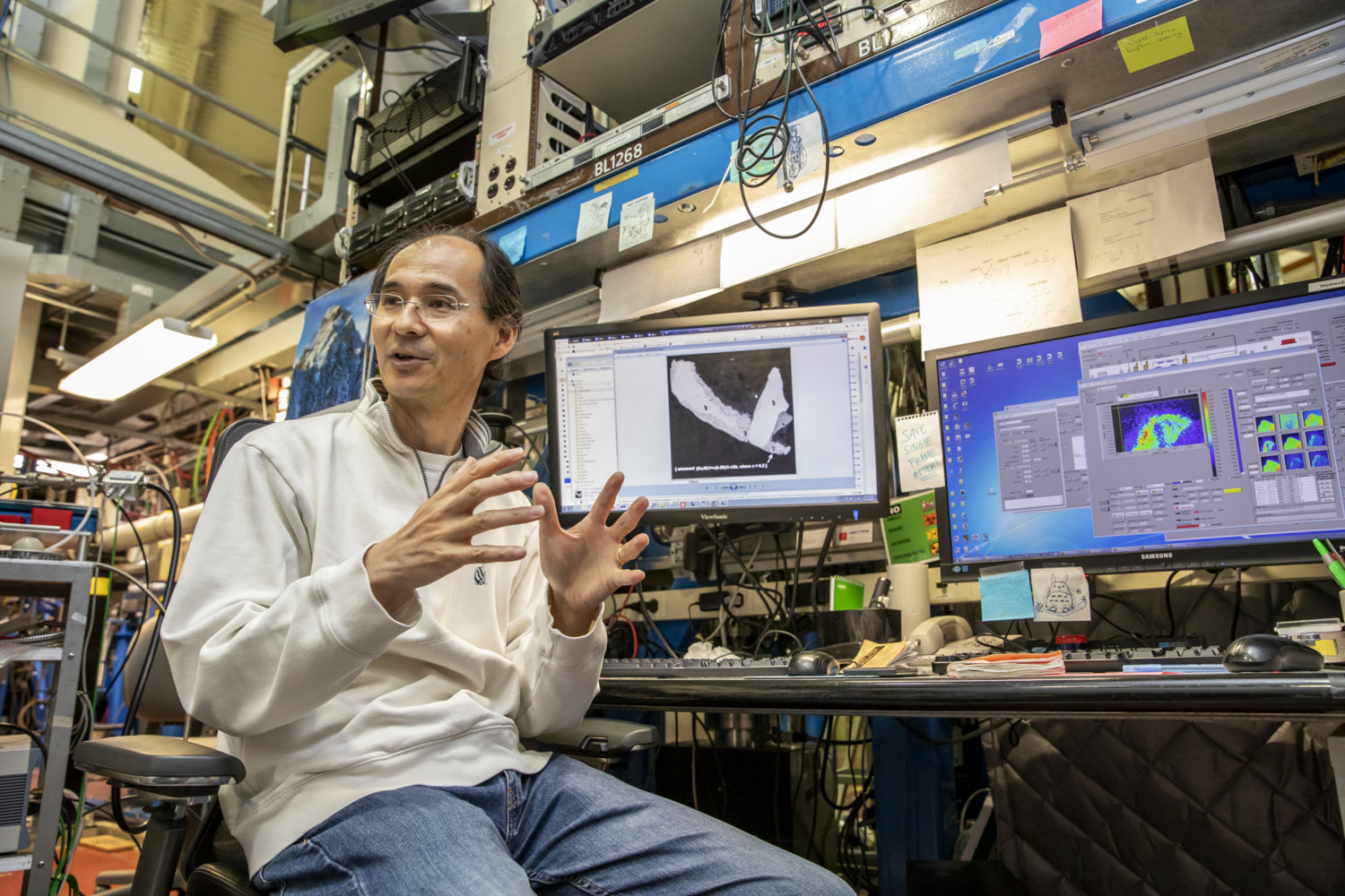
Nobumichi Tamura, a staff scientist at Berkeley Lab’s Advanced Light Source (ALS), studies a rare crystal sample at ALS Beamline 12.3.2. An X-ray technique at this beamline was key in a study that helped to confirm the discovery of the mineral ognitite. (Credit: Marilyn Chung/Berkeley Lab)
Like a tiny needle in a sprawling hayfield, a single crystal grain measuring just tens of millionths of a meter – found in a borehole sample drilled in Central Siberia – had an unexpected chemical makeup.
And a specialized X-ray technique in use at the Department of Energy’s Lawrence Berkeley National Laboratory (Berkeley Lab) confirmed the sample’s uniqueness and paved the way for its formal recognition as a newly discovered mineral: ognitite.
Based on this success with the technique at Berkeley Lab’s Advanced Light Source (ALS), the research team is employing it to study other tiny samples of promising candidates for new mineral discoveries. The ALS is a synchrotron that produces X-rays and other types of light for dozens of simultaneous experiments.
“The difficulty is that these minerals can be extremely rare and are only available in very small amounts,” said Nobumichi Tamura, a staff scientist at the ALS who helped to customize the experimental technique – known as X-ray Laue microdiffraction (and also micro-Laue X-ray diffraction) – to study tiny crystal samples including minerals. Tamura participated in the ognitite discovery and is now working with the same team to explore other samples.
Taking on the ‘desperate cases’
The ognitite mineral’s structure and other properties are detailed in a study published in May in Mineralogical Magazine and also documented in the European Journal of Mineralogy. The study also describes a new, cobalt-rich mineral variety – described as “cobaltian maucherite” – that Tamura explored using the same technique at the ALS.
“We are looking at cases where no conventional techniques can work,” Tamura said. “These are the desperate cases.”
He added, “I had been interested for years in developing this technique specifically to identify new minerals, because occasionally there are researchers who have an unknown material that they cannot resolve using any of the more conventional techniques.” In the cases of ognitite and the cobaltian maucherite, there are only individual samples of each that have been identified, to date.
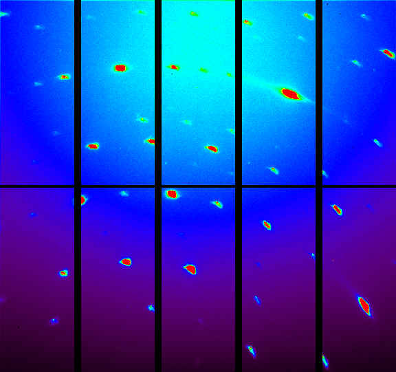
This image shows a diffraction pattern for the ognitite sample studied at Berkeley Lab’s Advanced Light Source. The pattern was obtained using a technique known as X-ray Laue microdiffraction. (Image courtesy of Nobumichi Tamura/Berkeley Lab)
The form of X-ray Laue microdiffraction employed at the ALS uses a narrowly focused X-ray beam that spans a range of energies to explore the atomic structure of materials in exquisite detail. The beam is focused to about a hundredth the diameter of a human hair.
Conventional single-crystal X-ray diffraction typically rotates crystal samples in an X-ray beam at a specific energy to help resolve their atomic structure, Tamura noted.
When crystal samples are so precious and small that researchers cannot easily extract them from surrounding materials without damaging the crystals, techniques including electron diffraction, single-crystal X-ray diffraction, and powder X-ray diffraction are typically out of the question.
The ALS technique, meanwhile, scans across the entire sample without the need to rotate the crystal, separate it from its surroundings, or prepare it any other way for study.
The entire scan is completed within a few minutes, though the data analysis for this technique is far more complex than for conventional diffraction and requires substantial computing power. Researchers use computer clusters at Berkeley Lab’s National Energy Research Scientific Computing Center (NERSC) and Laboratory Research Computing to process the data from the Laue microdiffraction experiments.
Catherine Dejoie, now a beamline scientist at the European Synchrotron Radiation Facility (ESRF), was hired as an ALS postdoctoral researcher in 2009 specifically to develop a method for analyzing the data from the Laue microdiffraction technique to resolve the atomic structure of materials. She worked in close collaboration with Tamura.
Chemical clues in tiny sample
Andrei Barkov, director of the Research Laboratory of Industrial and Ore Mineralogy at Cherepovets State University in Russia, led the international team credited with the ognitite discovery and was the lead author of the ognitite study.
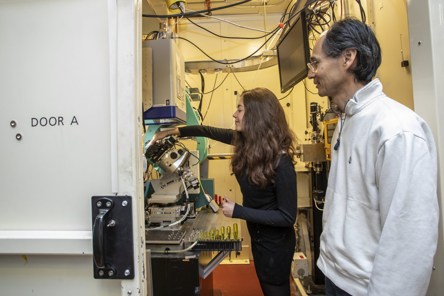
Elise Grenot, a student researcher from ENSTA, an engineering school in France, prepares a mineral sample for study using X-rays at Berkeley Lab’s Advanced Light Source. Nobumichi Tamura, an ALS staff scientist who participated in a study that helped to confirm ognitite as a new mineral, is pictured at right. (Credit: Marilyn Chung/Berkeley Lab)
That team included Tamura and Camelia Stan – Stan was a researcher at the ALS who participated in the ognitite study but has since left Berkeley Lab. Elise Grenot, a student researcher from France’s École Nationale Supérieure de Techniques Avancées (ENSTA), an engineering school, is now assisting Tamura with the latest round of candidate new-mineral experiments at the ALS.
Barkov learned about the technique developed at Berkeley Lab through his connection to Björn Winkler, a professor at Goethe University Frankfurt in Germany who was familiar with the ALS technique.
Barkov had already participated in several other successful mineral discoveries, including studies that led to the formal recognition of tatyanaite, edgarite, laflammeite, and menshikovite as new minerals. But the sample now known as ognitite was challenging to confirm as a new mineral although its chemistry appeared to be unique, Barkov noted.
“This mineral was suspected to be potentially new on the basis of its composition, which is unusually enriched in bismuth,” he said. “We could find just a single specimen, as a tiny grain. The grain is so small – that’s why the micro-Laue contributions from Nobu Tamura were so important.”
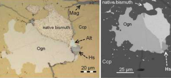
This reflected-light microphotograph, at left, shows the ognitite grain (Ogn), as well as bismuth, hessite (Hs), altaite (Alt), and magnetite (Mag). At right, a backscattered electron image also shows the mineral composition of the sample. (Credit: Mineralogical Magazine, May 8, 2019,
DOI: 10.1180/mgm.2019.31)
It took two attempts, including a follow-up round of experiments at the ALS for the second effort, to receive recognition of ognitite as a unique mineral from the Commission on New Minerals, Nomenclature and Classification of the International Mineralogical Association (IMA). The IMA reported 5,413 recognized minerals as of November 2018, and the list typically grows by 30 or more minerals each year after review and approval by the commission.
Ognitite contains nickel, bismuth, and tellurium. The study notes that its crystal structure is similar to a mineral called melonite, which is also composed of nickel and tellurium but is not associated with a high concentration of bismuth. And ognitite is chemically similar to the mineral tellurohauchecornite, which is composed of nickel, bismuth, tellurium, and sulfur.
New mineral is named for Ognit mineral complex in Siberia
Barkov said the ognitite discovery team’s first choice was to name it “baikalite” after Lake Baikal, which is in the region where the new mineral was discovered, but this name was not approved by the IMA. The commission instead favored “ognitite” as the mineral find was sourced from a place known as the Ognit ultramafic complex in Siberia’s Sayan Mountains region.
This geological formation is known to be rich in metal deposits, including rare platinum-group elements, nickel, and chromium.
The cobaltian maucherite sample was recovered from nickel-rich arsenides in the same Ognit complex, Barkov said, and measured just 20 millionths of a meter across. Because of its size and rarity, “it could only be characterized structurally” using the micro-Laue technique, he said.
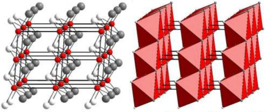
These diagrams show the atomic crystal structure of ognitite. At left, atoms in the crystalline structure are represented in red (nickel), white (tellurium), and gray (bismuth). At right, a polyhedral representation of the crystal structure. (Credit: Mineralogical Magazine, May 8, 2019, DOI: 10.1180/mgm.2019.31)
His team is exploring this type of formation in other parts of Russia, and the rock formations of particular interest can vary in size from about a kilometer to tens of kilometers, he said.
“We collect and examine, in detail, thousands of rock specimens and ore samples, and many more mineral grains,” he said. “As a result of these efforts, single grains of potentially new minerals may be found.”
His team typically uses optical microscopes, scanning electron microscopes, a technique known as energy-dispersive X-ray spectroscopy, wavelength-dispersive spectroscopy, and conventional X-ray diffraction to study mineral samples that have been collected over a span of decades.
From Russia to the ALS
Barkov contacted Björn Winkler to find out if he could create a synthetic form of ognitite, and also to synthesize other mineral samples.
“Professor Winkler has a solid background and proper facilities at his lab to synthesize new compounds that are analogous to potentially new minerals,” Barkov said. Winkler had already established a collaboration with Tamura, and Barkov then reached out to Tamura about the possibility of studying the ognitite sample at the ALS.
Dejoie, who helped to develop the data analysis methods to support the use of the ALS technique for studying the structure of tiny crystals, has returned to the ALS nearly every year to conduct experiments using this technique, and to improve upon the data-analysis methods. She said that in her own research she is now using the technique for time-resolved experiments that track how materials transition from one state of matter to another.
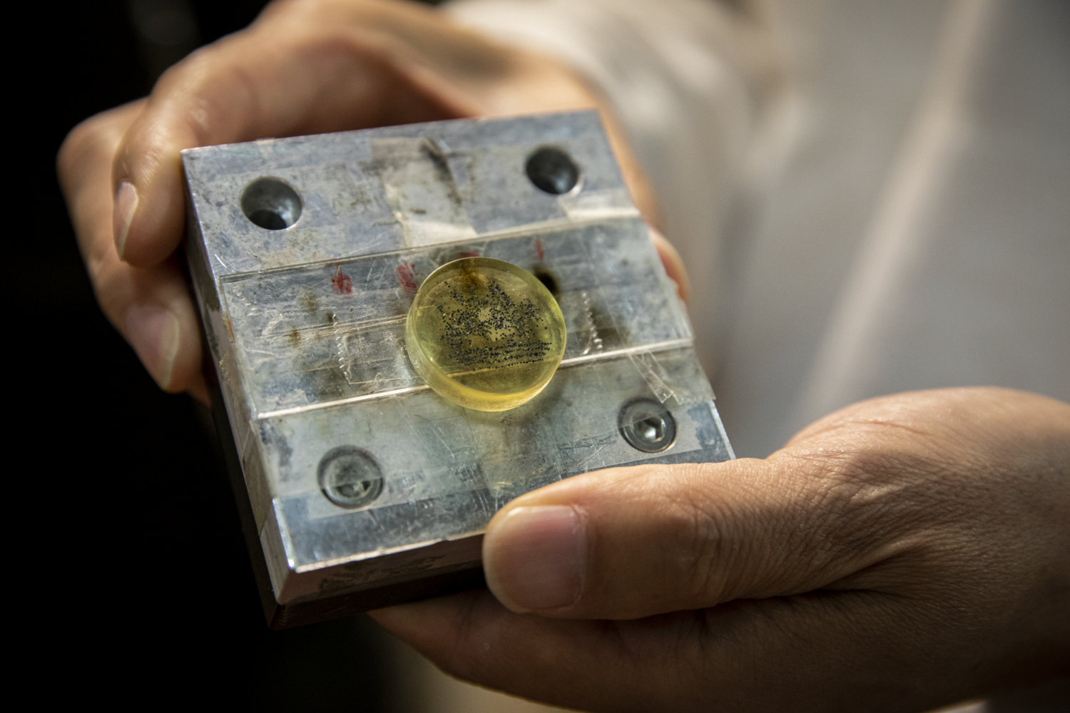
Nobumichi Tamura, a staff scientist at Berkeley Lab’s Advanced Light Source, holds an X-ray sample platform with an epoxy disk (center) encapsulating mineral samples. (Credit: Marilyn Chung/Berkeley Lab)
While X-ray Laue microdiffraction is not unique among the synchrotron light sources of the world, Dejoie and Tamura noted that its specialized application at the ALS and the maturity of its data-analysis methods are unique.
“We started to look at really small crystals – crystals that you cannot look at with a classic setup,” Dejoie recalled.
Growing interest
She noted that the technique can be used to resolve the timing of processes such as chemical reactions and structural changes in materials.
The Laue microdiffraction technique that she worked on at the ALS “is a really interesting alternative to electron diffraction,” Dejoie said, or at least a complementary tool for studying crystal structure, as it can quickly gather an entire high-precision data set.
She noted that an adaptation of Laue microdiffraction could also be useful for crystal studies at light sources known as X-ray free-electron lasers (XFELs), which have ultrashort, bright pulses.
“It’s funny to see the parallel – we were already using a similar kind of approach” to characterize the structure of crystals in a single pass, and without the need to rotate them or orient them in a particular way, before this was tried in XFEL studies.
In an XFEL technique known as “serial crystallography,” many crystal samples are streamed into the path of narrow-energy X-ray pulses. In these experiments, information is gathered from individual X-ray pulses striking randomly oriented crystals of the same sample type to develop a comprehensive 3D atomic structure.
Dejoie served as the lead author of a 2015 study detailing how the Laue diffraction technique of using a broad-energy X-pulse to strike single or multiple randomly oriented crystals simultaneously could be adapted for use at XFELs as a new “snapshot” approach to conventional serial crystallography.
It is gratifying, she said, to learn that the synchrotron-based technique for Laue microdiffraction she worked to develop at the ALS was helpful in confirming a new mineral. “It’s always good when you see something you’ve been working on getting some interest. It means it’s spreading, and that there may be a bit more development and more people working on it.”
The ALS and NERSC are both DOE Office of Science User Facilities.
The team participating in the ognitite discovery also included researchers from the University off Florence in Italy, Siberian Federal University in Russia, McGill University in Canada, and The Natural History Museum in the U.K. The ALS is supported by the DOE Office of Basic Energy Science. Individuals participating in the study were supported, in part, by the Russian Foundation for Basic Research and the U.K.’s Natural Environment Research Council.
###
Founded in 1931 on the belief that the biggest scientific challenges are best addressed by teams, Lawrence Berkeley National Laboratory and its scientists have been recognized with 13 Nobel Prizes. Today, Berkeley Lab researchers develop sustainable energy and environmental solutions, create useful new materials, advance the frontiers of computing, and probe the mysteries of life, matter, and the universe. Scientists from around the world rely on the Lab’s facilities for their own discovery science. Berkeley Lab is a multiprogram national laboratory, managed by the University of California for the U.S. Department of Energy’s Office of Science.
The Office of Science is the single largest supporter of basic research in the physical sciences in the United States, and is working to address some of the most pressing challenges of our time. For more information, please visit science.energy.gov.