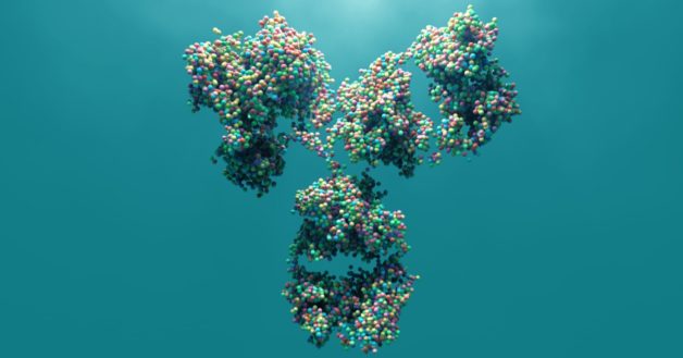
Vaccines, which help the body recognize infectious microorganisms and stage a stronger and faster response, are made up of proteins that are specific to each type of microorganism. In the case of a virus, viral proteins – or antigens – can sometimes be attached to a protein scaffold to help mimic the shape of the virus and elicit a stronger immune response. Using scaffolds to approximate the natural configuration of the antigen is an emerging approach to vaccine design.
A team of scientists led by David Baker at the University of Washington developed a method to design artificial proteins to serve as a framework for the viral antigens. Their study was published recently in the journal eLife. Berkeley Lab scientists collected data at the Advanced Light Source to visualize the atomic structure and determine the dynamics of the designed scaffolds.
“When bound, the scaffolds assume predicted geometries, which more closely approximate the virus shape and thereby maximize the immune response,” said Banu Sankaran, a research scientist in the Molecular Biophysics and Integrated Bioimaging (MBIB) Division. “It was exciting to collaborate on this method to predictably design frameworks, which could lead to more effective vaccines, especially for viruses that we don’t have a scaffold for.”
The team also included Peter Zwart, MBIB staff scientist; small angle X-ray scattering data were collected at the SIBYLS beamline by MBIB’s Kathryn Burnett and Greg Hura.
The Advanced Light Source is a DOE Office of Science user facility.
Media Contact:
[email protected]
510-486-5183

