
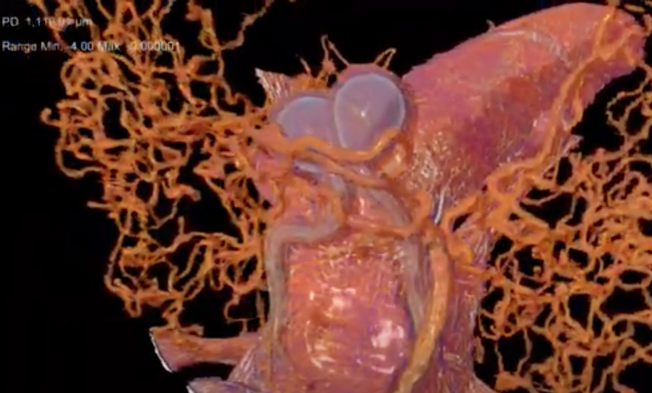
An animation produced from an X-ray microtomography scan of a female tsetse fly’s abdomen. The animation highlights the female reproductive system, including a milk gland used to feed developing larvae. (Credit: Geoffrey Attardo/UC Davis)
To better understand the unique reproductive biology of tsetse flies, which are carriers of the parasites that cause a deadly infection known as African sleeping sickness, researchers explored the intact organs and tissues of tsetse flies using a powerful 3D X-ray imaging technique at Berkeley Lab.
The imaging technique provided new insights into how the flies’ specialized biology governs mating and reproductive processes, including female flies’ unique lactation and their delivery of a single fully developed larvae per birthing cycle – whereas most other insect species lay eggs. The ALS produces X-rays and other forms of light for a broad range of simultaneous scientific experiments.
Fly samples were prepared at various stages of the reproductive cycle, and researchers are ultimately aiming to couple the imaging data with gene expression and biochemical data from the same stages of this cycle.
“We want to understand what changes are happening during this process, how the process is being mediated, and if it can be manipulated to artificially repress females in the wild from mating,” said Geoffrey Attardo, an assistant professor of entomology and nematology at UC Davis – which would ideally curb disease transmission. In 2015, about 3,500 people died from African sleeping sickness, and about 11,000 people are now estimated to be infected. The infection is fatal if it is left untreated by medication.
Attardo led a study, published in the journal Insects, detailing the ALS imaging work. The ALS experiments yielded better results than expected, he said.
While some other techniques require dissection and staining processes that introduce damage to the delicate samples, “This project allowed us to create a detailed 3D visualization of the reproductive tissues in their native context, with minimal experimental manipulation,” he said.
The ALS experiments provided a first, detailed look at the intact structure of a sperm-delivery structure called a spermatophore that fully occupies a female fly’s uterus after mating, for example, and detailed imaging of other reproductive-tract tissues relevant to lactation and birth.
“I love that as a staff member at the ALS, I can help enable science that makes a difference in the world,” said Dula Parkinson, an ALS staff scientist and Diffraction and Imaging Program leader who participated in the study.