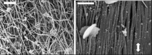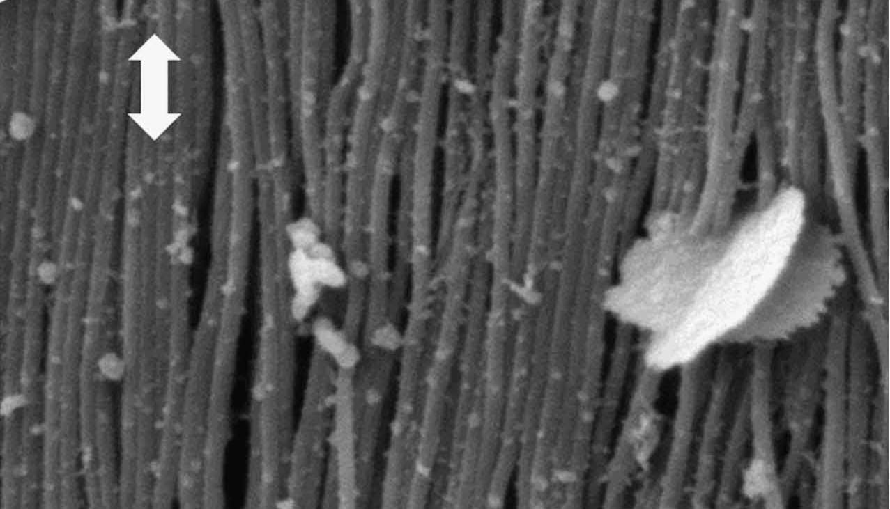
(Left) Collagen fibrils in the dermis of the skin are normally curvy and highly disordered, but (right) in response to a tear align themselves with the tension axis (arrow) to resist further damage.
When weighing the pluses and minuses of your skin add this to the plus column: Your skin – like that of all vertebrates – is remarkably resistant to tearing. Now, a collaboration of researchers at the U.S. Department of Energy (DOE)’s Lawrence Berkeley National Laboratory (Berkeley Lab) and the University of California (UC) San Diego has shown why.
Making good use of the X-ray beams at Berkeley Lab’s Advanced Light Source (ALS), the collaboration made the first direct observations of the micro-scale mechanisms behind the ability of skin to resist tearing. They identified four specific mechanisms in collagen, the main structural protein in skin tissue, that act synergistically to diminish the effects of stress.
“Collagen fibrils and fibers rotate, straighten, stretch and slide to carry load and reduce the stresses at the tip of any tear in the skin,” says Robert Ritchie of Berkeley Lab’s Materials Sciences Division, co-leader of this study along with UC San Diego’s Marc Meyers. “The movement of the collagen acts to effectively diminish stress concentrations associated with any hole, notch or tear.”
Ritchie and Meyers are the corresponding authors of a paper in Nature Communications that describes this study. The paper is titled “On the tear resistance of skin.” The other authors are Wen Yang, Vincent Sherman, Bernd Gludovatz, Eric Schaible and Polite Stewart.

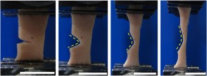
Sequence of images showing how skin when torn does not propagate but progressively yawns open under tensile loading.
Who among us does not pay close attention to the condition of our skin? While our main concern might be appearance, skin serves a multitude of vital purposes including protection from the environment, temperature regulation and thermal energy collection. The skin also serves as a host for embedded sensors.
Skin consists of three layers – the epidermis, dermis and endodermis. Mechanical properties are largely determined in the dermis, which is the thickest layer and is made up primarily of collagen and elastin proteins. Collagen provides for mechanical resistance to extension, while elastin allows for deformation in response to low strains.
Studies of the skin’s mechanical properties date back to 1831 when a physician investigated stabbing wounds that the victim claimed were self-inflicted. This eventually led to scientists characterizing skin as a nonlinear-elastic material with low strain-rate sensitivity. In recent years research has focused on collagen deformation but with little attention paid to tearing even though skin has superior tear-resistance to other natural materials.
“Our study is the first to model and directly observe in real time the micro-scale behavior of the collagen fibrils associated with the skin’s remarkable tear resistance,” Ritchie says.
Ritchie, Meyers and their colleagues performed mechanical and structural characterizations during in situ tension-loading in the dermis. They did this through a combination of images obtained via ultrahigh-resolution scanning and transmission electron microscopy, and small-angle X-ray scattering (SAXS) at ALS beamline 7.3.3.
“The method of SAXS that can be done at ALS beamline 7.3.3 is ideal for identifying strains in the collagen fibrils and watching how they move,” Ritchie says.
The research team began their work by establishing that a tear in the skin does not propagate or induce fracture, as does damage to bone or tooth dentin, which are comprised of mineralized collagen fibrils. What they demonstrated instead is that the tearing or notching of skin triggers structural changes in the collagen fibrils of the dermis layer to reduce the stress concentration. Initially, these collagen fibrils are curvy and highly disordered. However, in response to a tear they rearrange themselves towards the tensile-loading direction, with rotation, straightening, stretching, sliding and delamination prior to fracturing.
“The rotation mechanisms recruit collagen fibrils into alignment with the tension axis at which they are maximally strong or can accommodate shape change,” says Meyers. “Straightening and stretching allow the uptake of strain without much stress increase, and sliding allows more energy dissipation during inelastic deformation. This reorganization of the fibrils is responsible for blunting the stress at the tips of tears and notches.”
This new study of the skin is part of an on-going collaboration between Ritchie and Meyers to examine how nature achieves specific properties through architecture and structure.
“Natural inspiration is a powerful motivation to develop new synthetic materials with unique properties,” Ritchie says. “For example, the mechanistic understanding we’ve identified in skin could be applied to the improvement of artificial skin, or to the development of thin film polymers for applications such as flexible electronics.”
This research was primarily supported by the Air Force Office of Scientific Research. Berkeley Lab’s Advanced Light Source is a DOE Office of Science User Facility.
Additional Information
For more about the research of Robert Ritchie go here
For more about the research of Marc Meyers go here
For more about ALS Beamline 7.3.3 go here
# # #
Lawrence Berkeley National Laboratory addresses the world’s most urgent scientific challenges by advancing sustainable energy, protecting human health, creating new materials, and revealing the origin and fate of the universe. Founded in 1931, Berkeley Lab’s scientific expertise has been recognized with 13 Nobel prizes. The University of California manages Berkeley Lab for the U.S. Department of Energy’s Office of Science. For more, visit www.lbl.gov.
DOE’s Office of Science is the single largest supporter of basic research in the physical sciences in the United States, and is working to address some of the most pressing challenges of our time. For more information, please visit the Office of Science website at science.energy.gov/.

