Berkeley Lab scientists are developing new ways to see the unseen. Here are seven imaging advances (recently reported in our News Center) that are helping to push science forward, from developing better batteries to peering inside cells to exploring the nature of the universe.
1. Seeing DNA nanostructures in 3-D
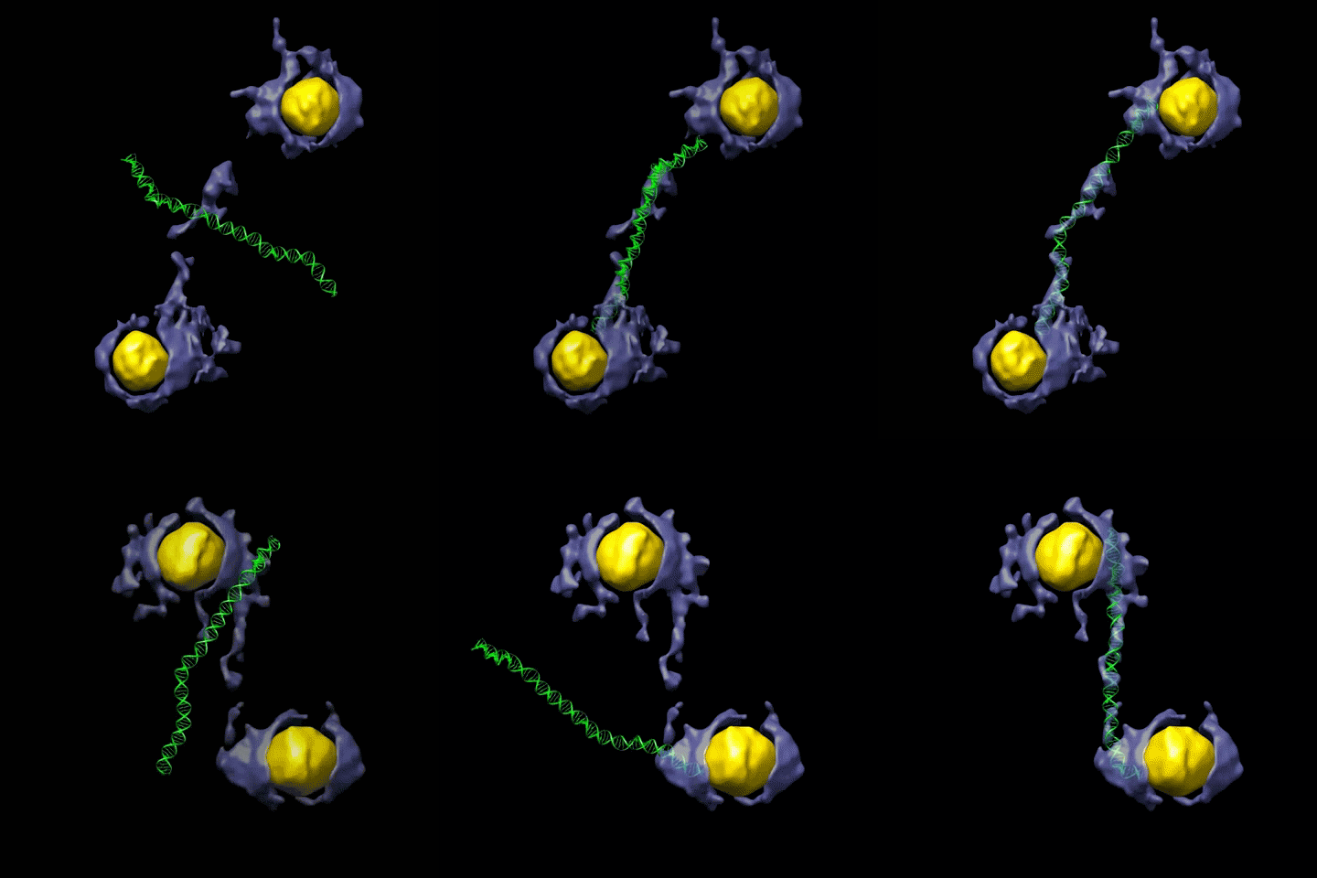
DNA segments can serve as a nanoscale building material, and scientists have devised a new way to see the shape of nanoscale DNA segments in 3-D. These images could help us understand how to attach DNA to other materials to build drug-delivery systems, and how to use it as a tiny marker for biological experiments. The images could also be used to map disease-relevant proteins in greater detail.
2. An ultrafast electron eye
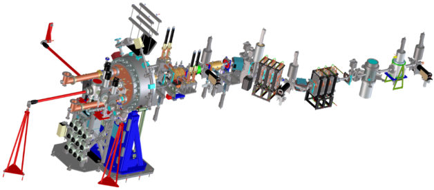
Scientists are using ultrafast pulses of highly focused electrons like a camera to study chemical processes and changes in materials at the atomic scale. How it works: An initial laser pulse triggers a reaction in a sample that is followed an instant later by an electron pulse to produce an image. A sequence of these images, which can capture processes that occur in just quadrillionths of a second, could provide new insight in how to make materials with custom properties and to enhance chemical reactions.
3. ‘Invisible’ elements unveiled
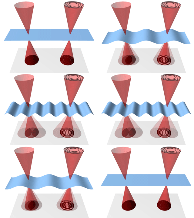
Electron microscopes can look at durable materials in atomic-scale detail, though more delicate materials pose a greater challenge. Recently, researchers figured out a way to modify a popular electron microscopy technique to look at a mix of materials, even those that would appear invisible to standard imaging techniques. The new method could be useful for viewing a mix of heavy and light materials such as battery components.
4. Redder is better

Just as sound is distorted to a lower pitch as an ambulance passes you with its siren blaring, light is shifted as objects move away from us. In space, the light from distant galaxies moving away from us is shifted to redder wavelengths. Special light sensors developed at Berkeley Lab, installed in a telescope-mounted camera, allow scientists to see better at redder wavelengths so they can capture the light from more distant galaxies. The sensitivity of this camera, called Mosaic-3, can detect objects billions of light years away.
5. DNA repair, time-lapse edition
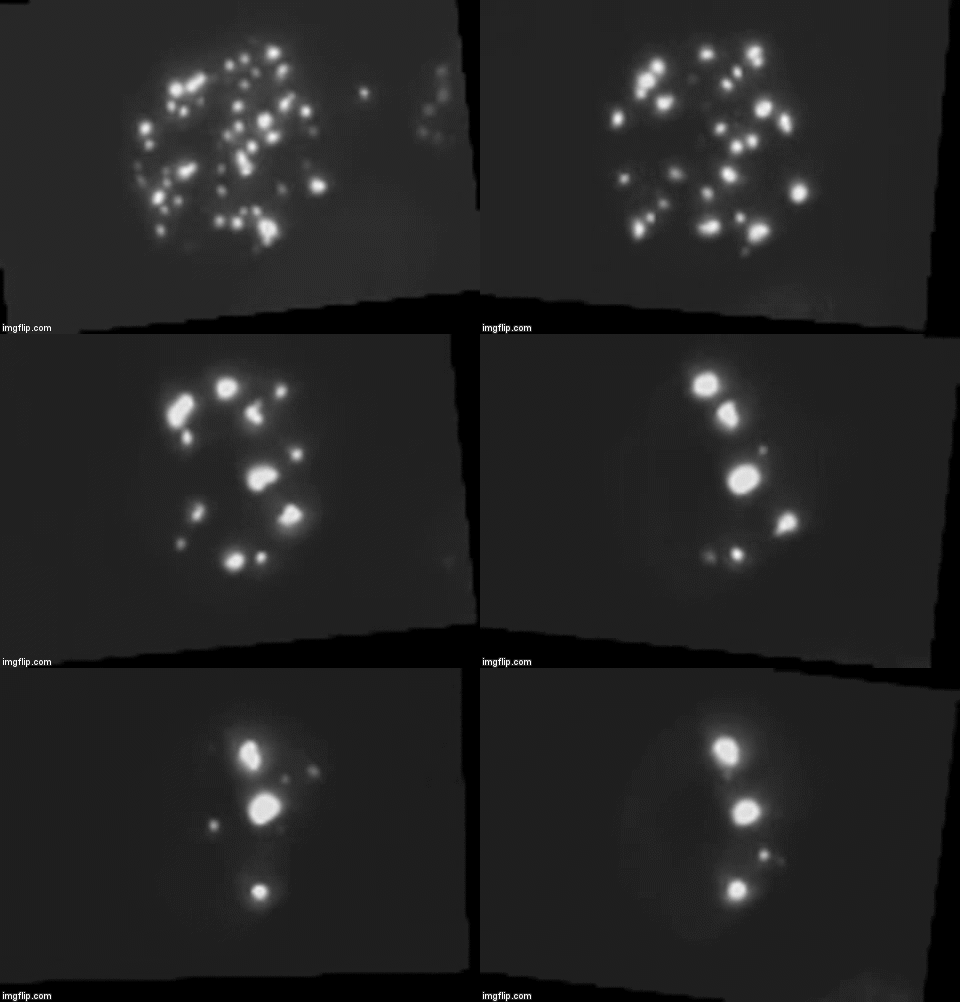
Time-lapse images can make complicated processes easier to understand. Our scientists developed a similar approach to measure DNA repair in human mammary epithelial cells. Their technique has shed light on how cells repair DNA strand breaks, which could help scientists learn how to protect astronauts from cosmic rays as well as refine radiotherapy protocols designed to kill tumors.
6. A 3-D journey inside the center of cells
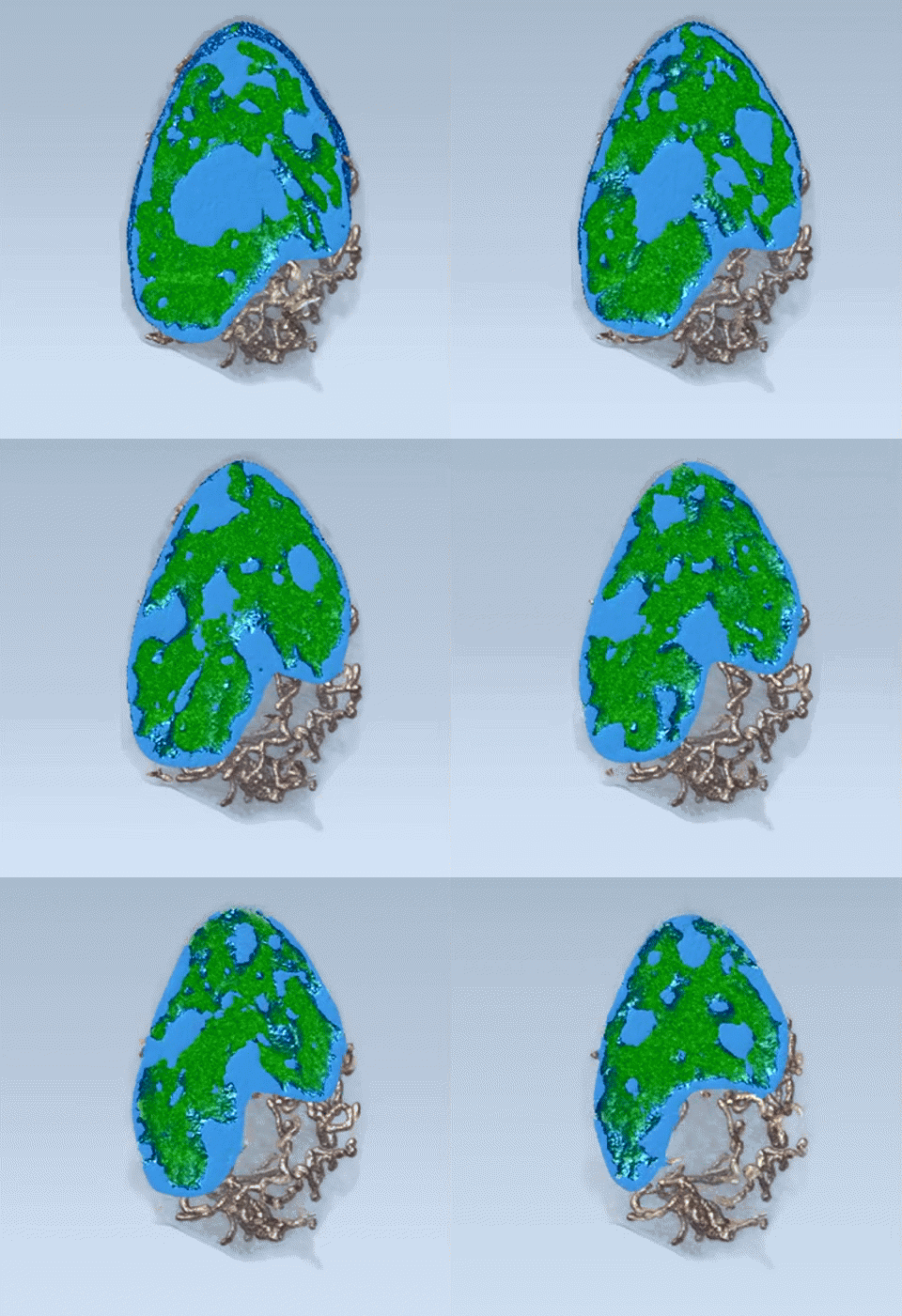
Detailed 3-D visualizations have enabled scientists to map the reorganization of genetic material that happens when a stem cell matures into a nerve. This animation shows a 3-D rendering of a nucleus in a mouse cell known as a “neuronal progenitor.” The view shown here slices from the surface of the nucleus through to its other side, and is color-coded for two types of genetic material: heterochromatin (blue) and euchromatin (green). This new imaging technique promises a better understanding of how genes are activated or silenced.
7. A window into longer lasting batteries
There’s a new tool in the push to engineer rechargeable batteries that last longer and charge more quickly. An X-ray microscopy technique recently developed at the Advanced Light Source, a DOE Office of Science User Facility, images nanoscale changes inside lithium-ion battery particles as they charge and discharge. Insights obtained from the imaging technique could help researchers improve batteries for electric vehicles as well as smart phones, laptops, and other devices.
