
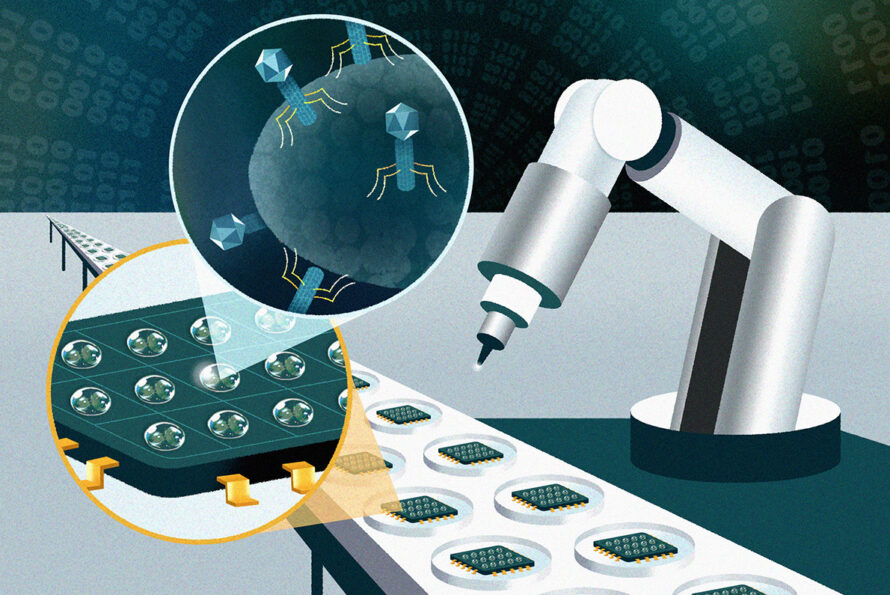
Combatting Antibiotic Resistance with Nanotechnology, Robotics, and AI
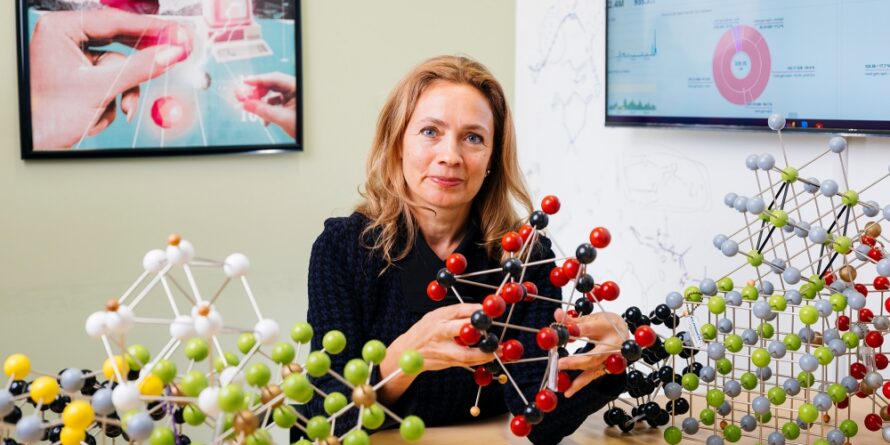
Berkeley Lab’s Kristin Persson Elected to the American Academy of Arts and Sciences
Three Berkeley Lab Researchers Named AAAS Fellows

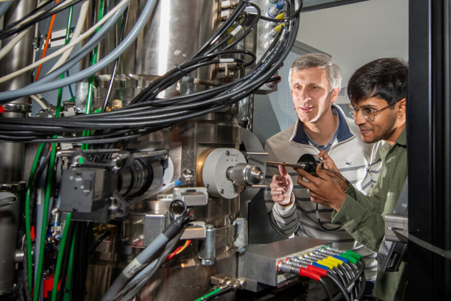
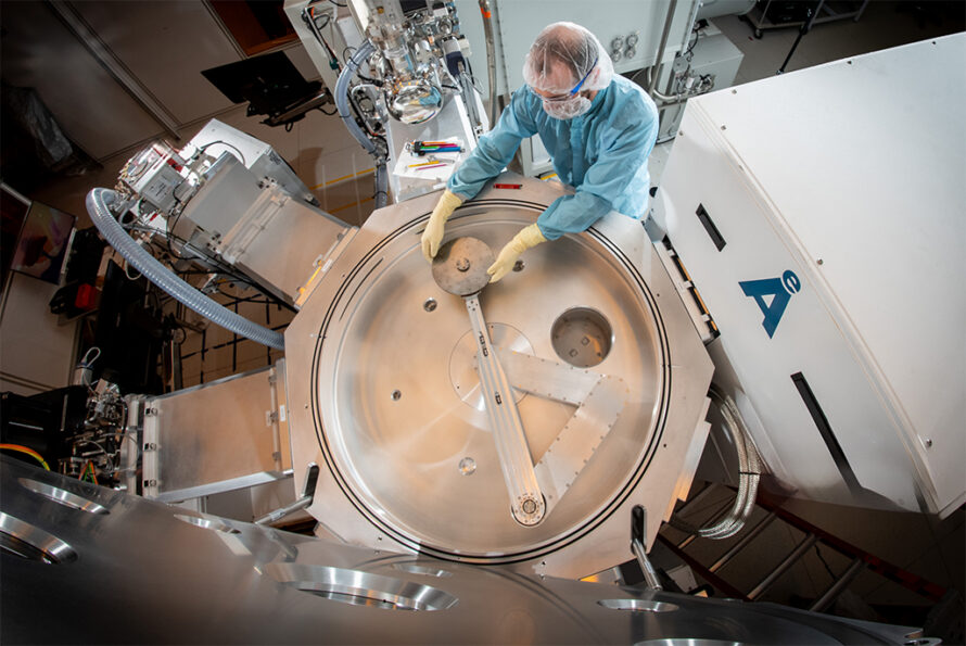
New Electron Microscopy Technique Reveals Atomic Structures From Nanocrystals Once Considered Unsolvable
Basics 2 Breakthroughs: Optimizing Materials for Next-Generation Microelectronics

Science on the Double: How an AI-Powered 'Digital Twin' Accelerates Chemistry and Materials Discoveries
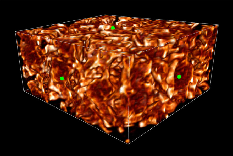
Crunching Big Data Into 3D Images Accelerates Discovery
A ‘Robot Pizza Chef’ Serving Up Better Quantum Computers
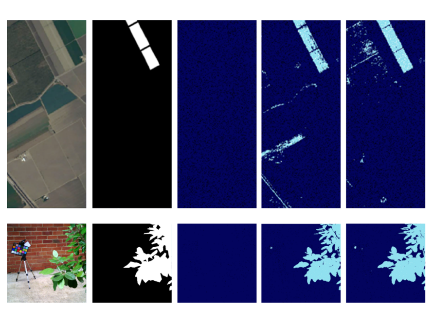
New AI Sensor ‘Sniffs’ Out Spectral Targets
Berkeley Lab Leads Effort to Build AI Assistant for Energy Materials Discovery

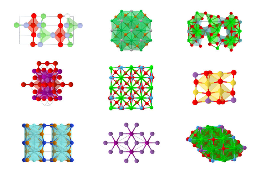
Accelerating Discovery: How the Materials Project Is Helping to Usher in the AI Revolution for Materials Science
From Nano to Nobel: National Lab Researchers Use MOFs to Solve Big Problems
