Atomic Neighborhoods in Semiconductors Provide New Avenue for Designing Microelectronics
Electron Microscopy Reveals New Method to Make Exotic Metal Alloys
Optimized Materials in a Flash
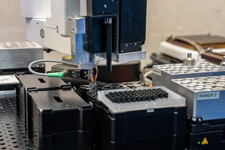
How AI and Automation are Speeding Up Science and Discovery
Atomic X-ray Laser Opens Door to Attosecond Imaging
Expert Interview: Ashfia Huq on the Macroscale Promise of Nanoscale Science

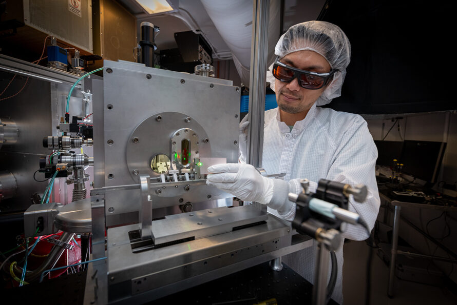
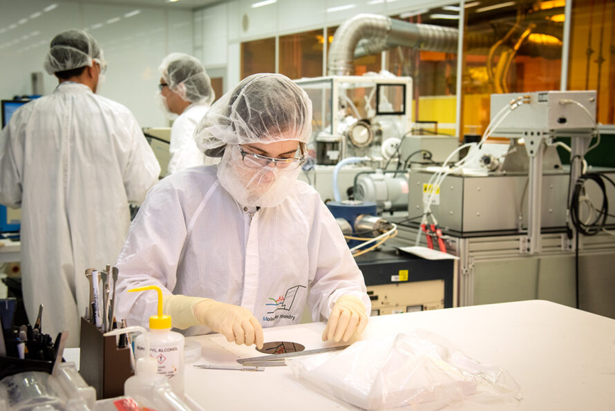
Six Scientific Advances Made Possible by Berkeley Lab’s Molecular Foundry
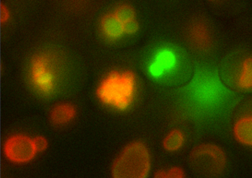
New Process Uses Microbes to Create Valuable Materials from Urine
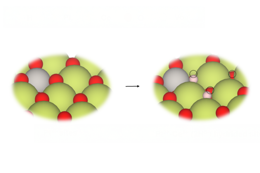
Catalysts Get a Boost with Atomic-Level Tinkering
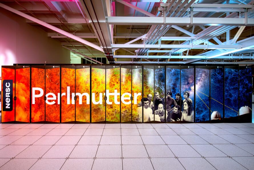
Five Ways Berkeley Lab's NERSC is Revolutionizing Scientific Research
Computational Chemistry Unlocked: A Record-Breaking Dataset to Train AI Models has Launched

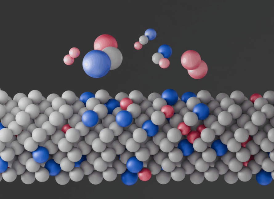
Scientists Crack Decades-Old Puzzle in CO2-to-Fuel Conversion