
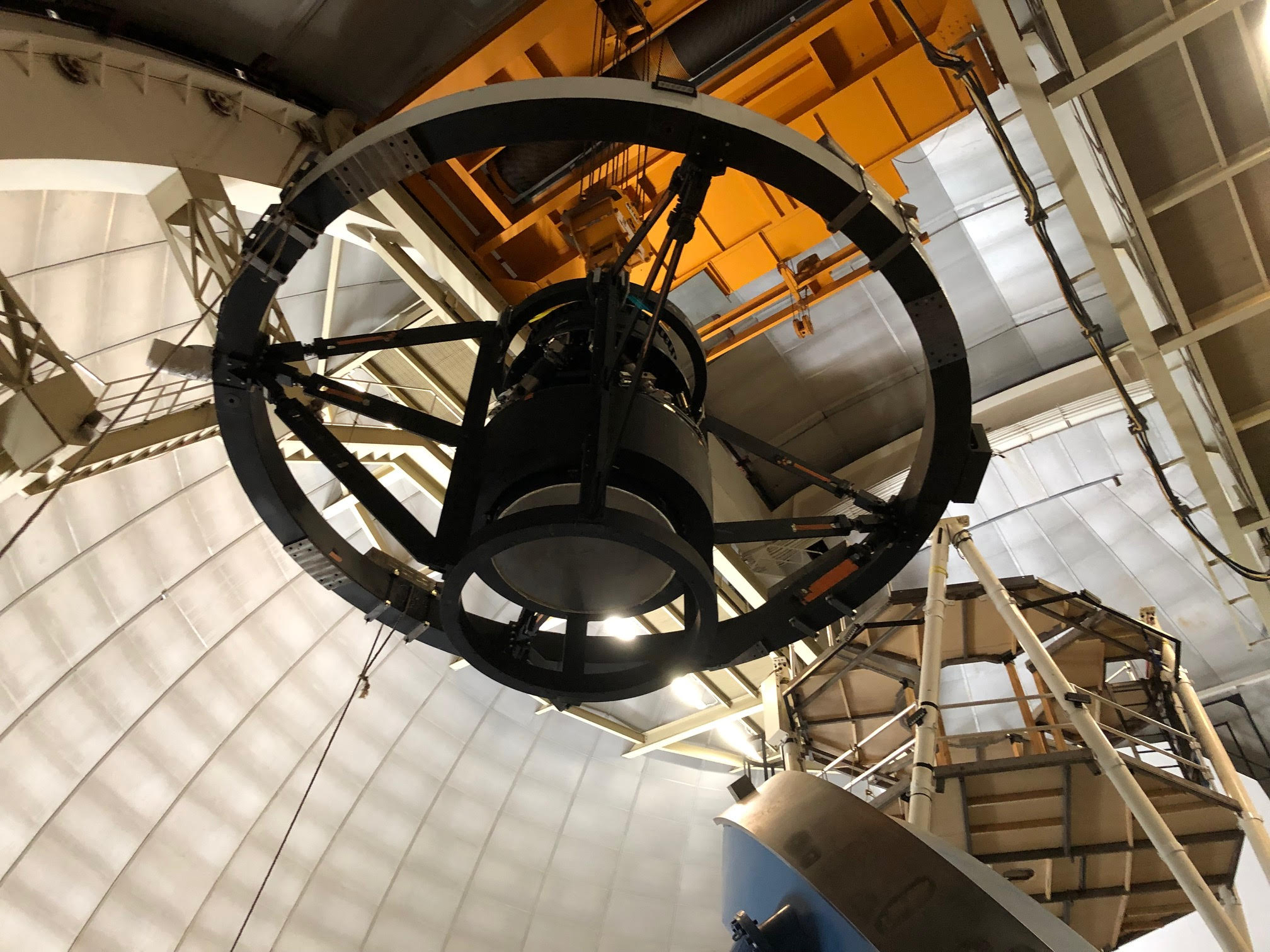
Topping Off a Telescope with New Tools to Explore Dark Energy
How Berkeley Lab is Advancing Microelectronics

Four College Teams to Converge at Berkeley Lab for DOE CyberForce Competition

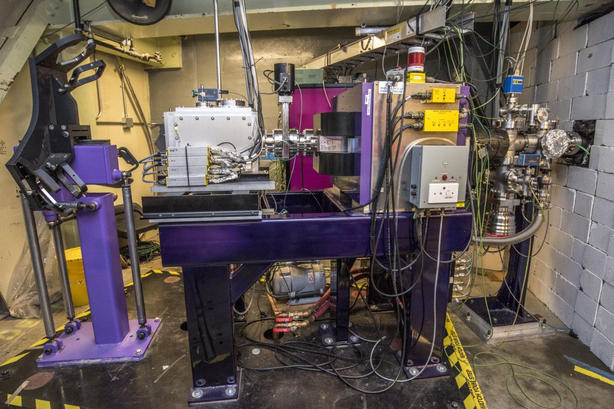
FIONA Measures the Mass Number of 2 Superheavy Elements: Moscovium and Nihonium
Potential New Way to Boost Biofuels and Bioproducts Production
Four Berkeley Lab Scientists Named AAAS Fellows

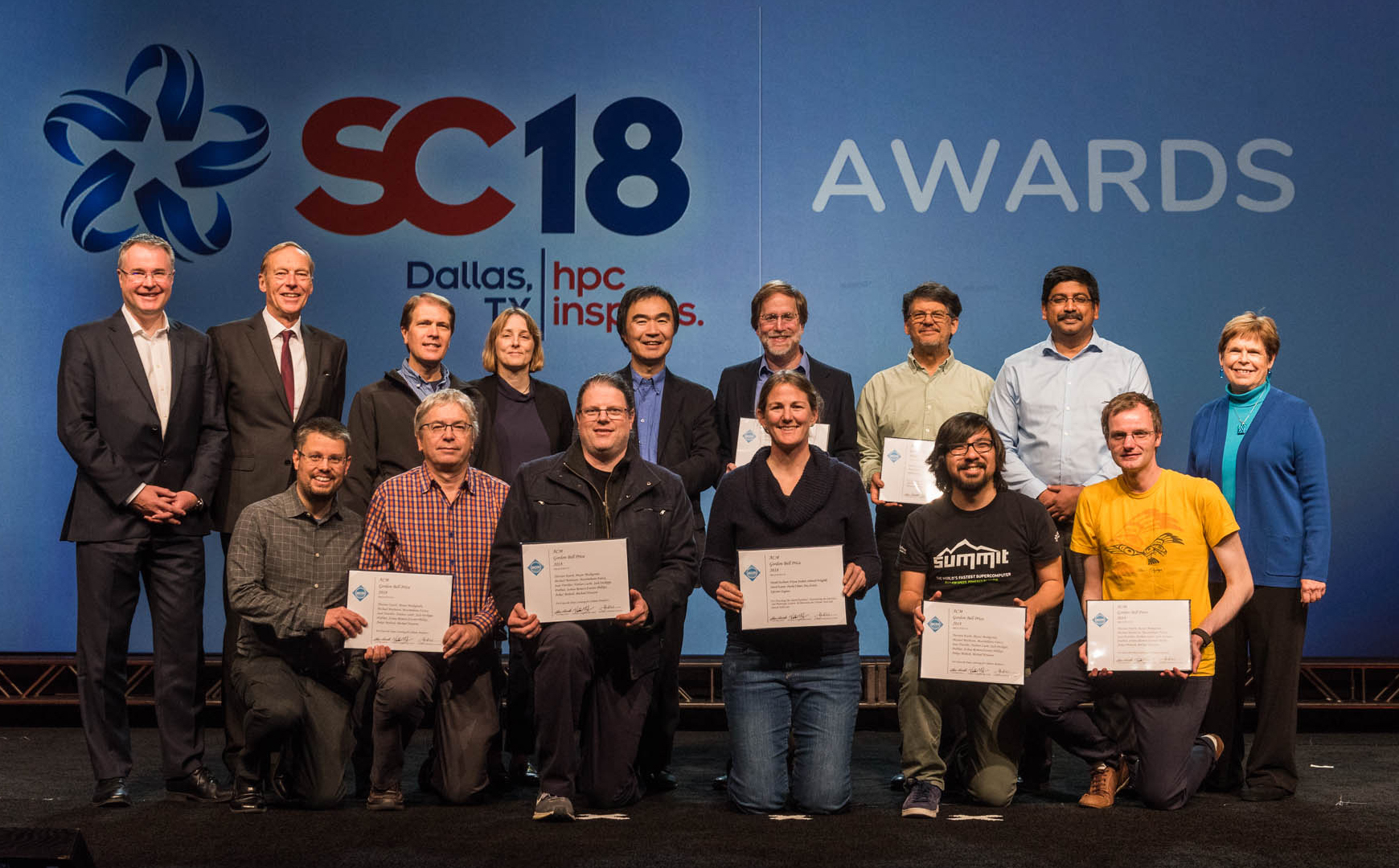
Berkeley Lab, Oak Ridge National Lab Share 2018 ACM Gordon Bell Prize
Berkeley Lab Wins 2018 R&D 100 Award; Special Recognition Award for Cyclotron Road Startup

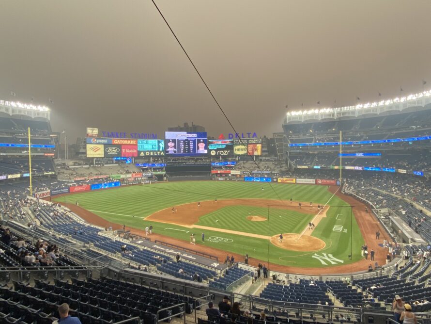
Q&A: How to Protect Yourself and Your Family From Wildfire Smoke
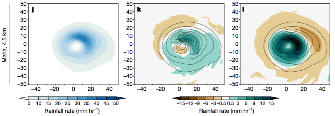
Climate Simulations Project Wetter, Windier Hurricanes
Scientists Bring Polymers Into Atomic-Scale Focus

Scientists Capture Photosynthesis in Unprecedented Detail
