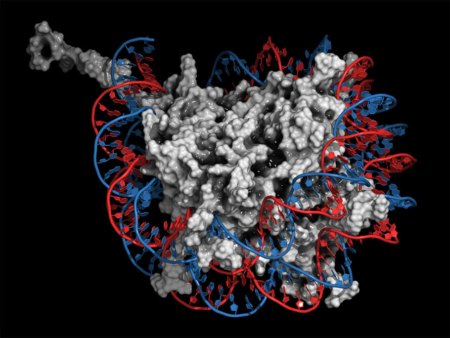
A Better Understanding of DNA Unpacking
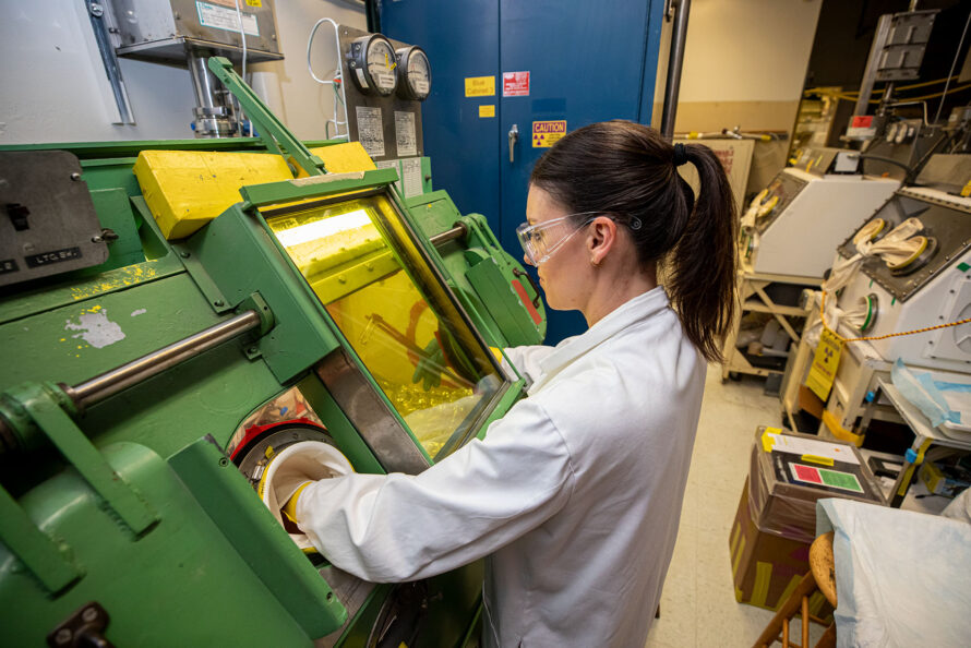
Caught in the Actinium
How to Make Sustainable Products Faster with Artificial Intelligence and Automation

Tracking Down Toxic Metals From Tobacco Smoke

Two Berkeley Lab Researchers Elected to the National Academy of Sciences

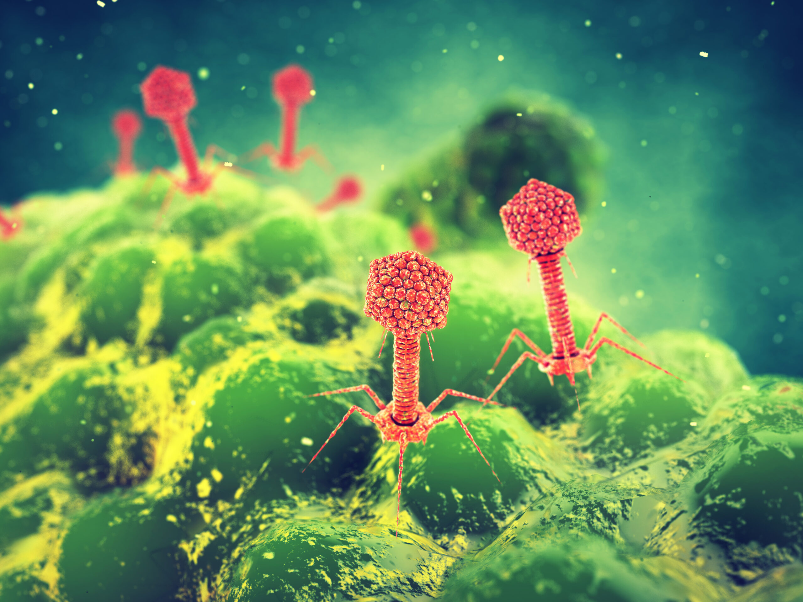
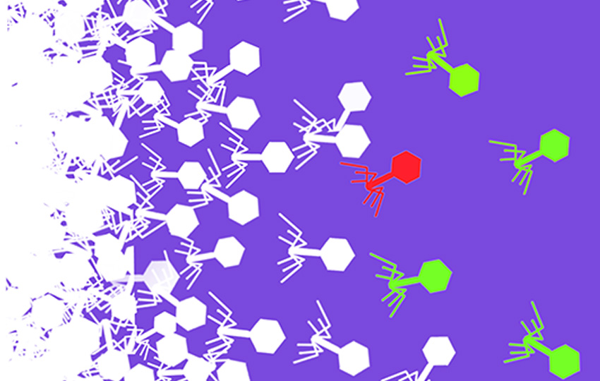
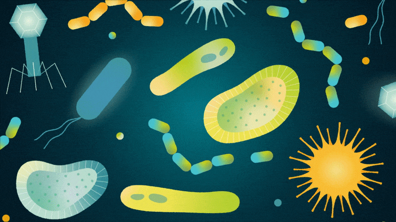
Barcoding Bacteriophages: New Method Could Unleash Powerful Biotechnology Applications
Berkeley Lab Launches Research Projects to Support National Biopreparedness and Response Efforts

Natural or Not? Scientists Aid in Quest to Identify Genetically Engineered Organisms
Carpets Retain a Stubborn Grip on Pollutants from Tobacco Smoke

Breaking Barriers in Drug Delivery with Better Lipid Nanoparticles

Eva Nogales Wins Shaw Prize in Life Science and Medicine

Seven Ways Berkeley Lab Researchers Improve Health for All