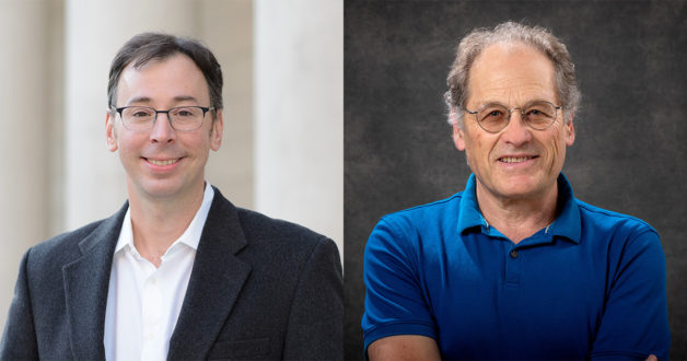
2 Berkeley Lab Physicists Elected into the National Academy of Sciences
How Venice, Italy Can Cut Carbon Emissions from Social Housing

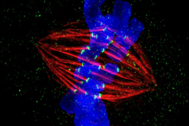
First ‘Telomere to Telomere’ Human Genome Reveals Secrets of the Centromere
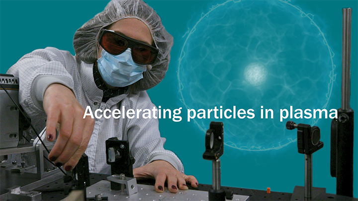
Basics 2 Breakthroughs
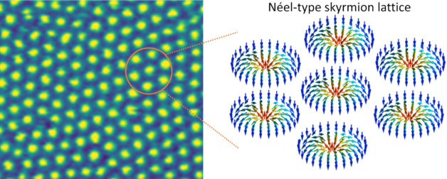
Skyrmions on the Rise – New 2D Material Advances Low-Power Computing
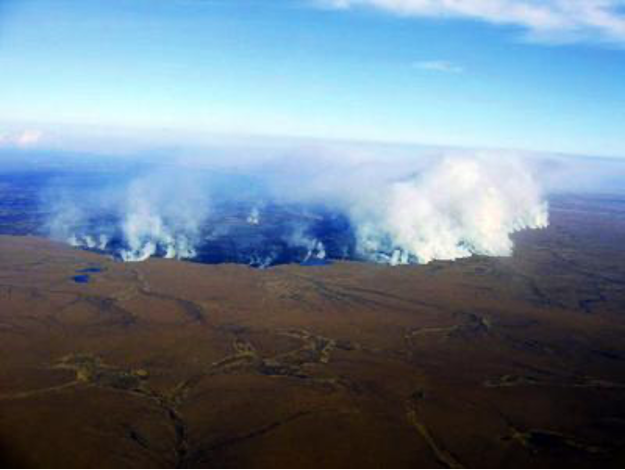
Microbial Response to a Changing and Fire-Prone Arctic Ecosystem
A Day in the Half-Life

Capturing Carbon With Inspiration From Battery Chemistry
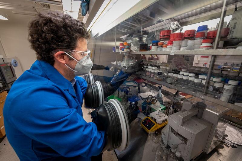
Using Hundred-Year-Old Chemistry to Capture Carbon Directly From Air
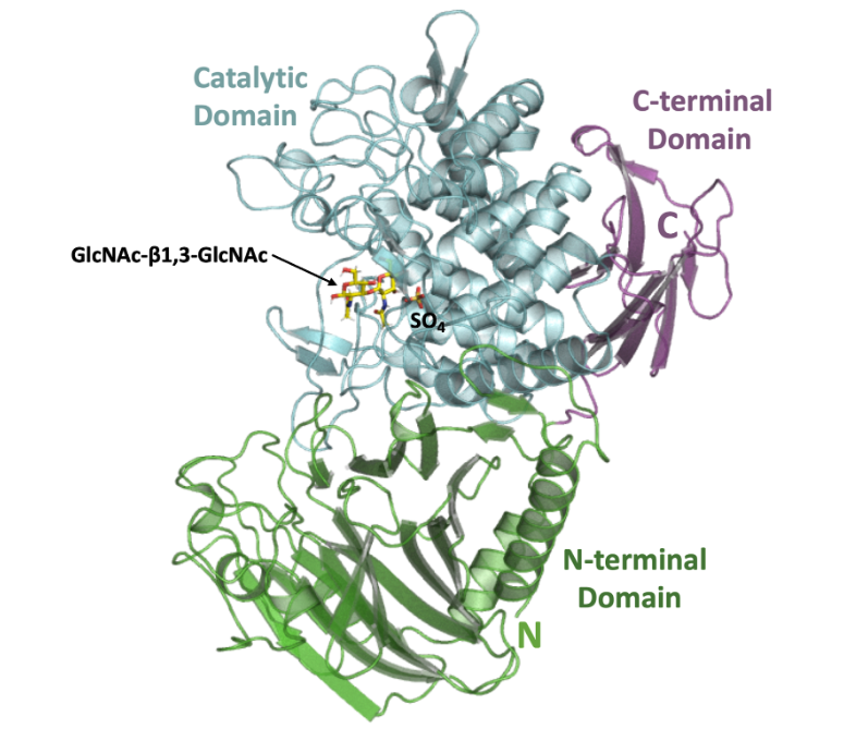
Newly Discovered Bacterial Enzyme Produces Useful Biopolymer
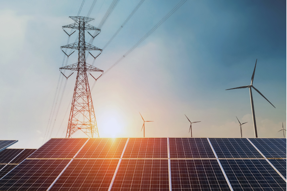
Record Amounts of Zero-carbon Electricity Generation and Storage Now Seeking Grid Interconnection
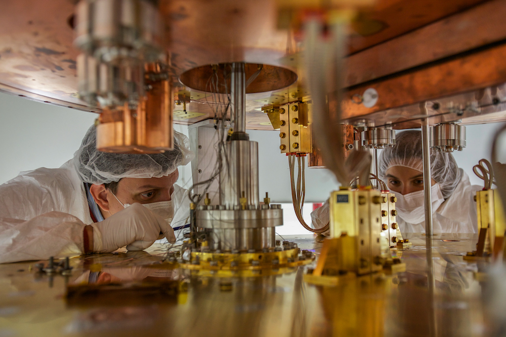
CUORE Team Places New Limits on the Bizarre Behavior of Neutrinos