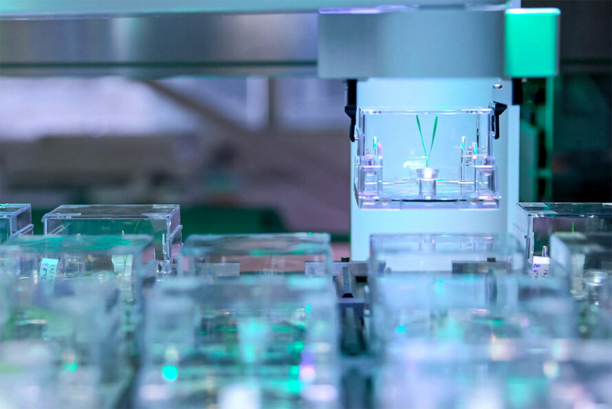
Meet EcoBOT: The Autonomous Lab Standardizing Plant-Microbe Research
EcoFABs Could Help Fuel AI in Agriculture
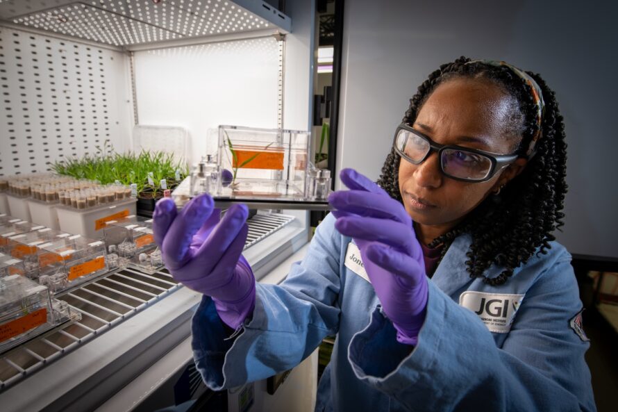
Advancing the Frontiers of Genetic Science at the Joint Genome Institute
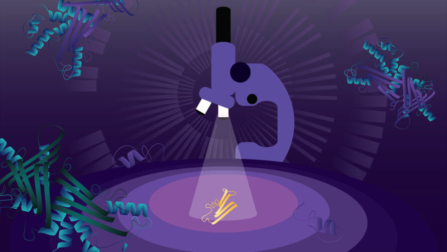
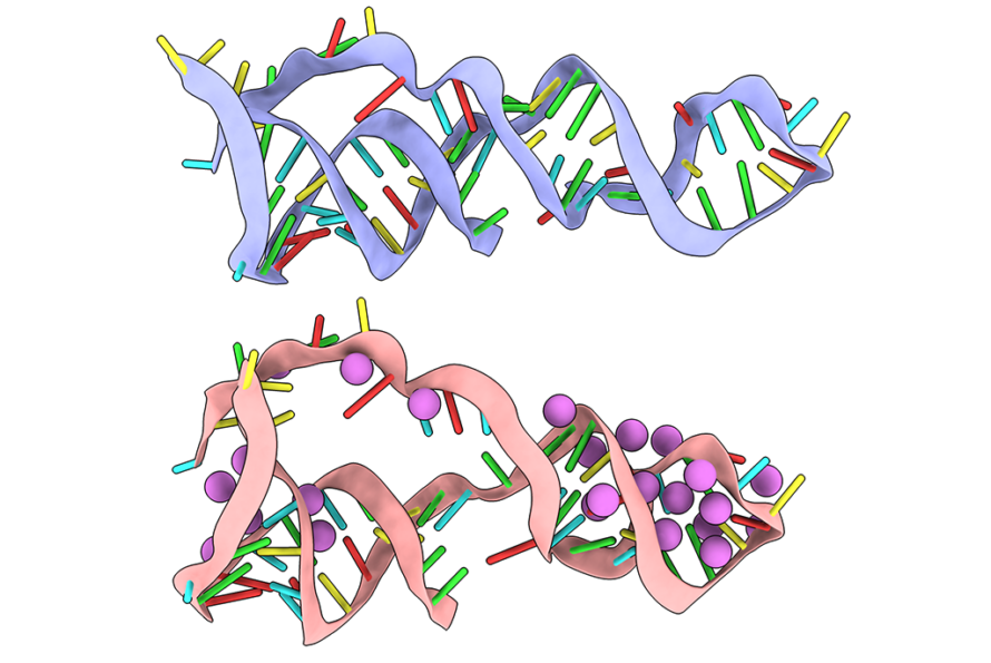
From Sequence to Structure: A Fast Track for RNA Modeling
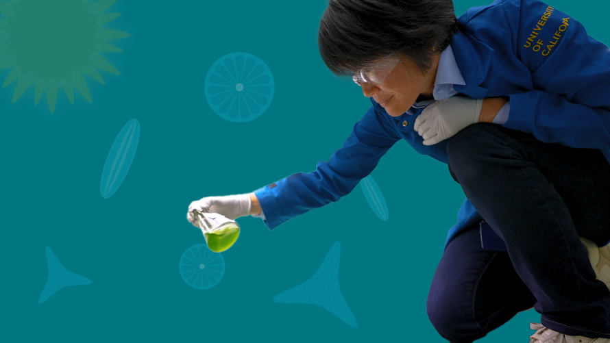
Sequencing the Mysterious Microbes of the San Francisco Estuary

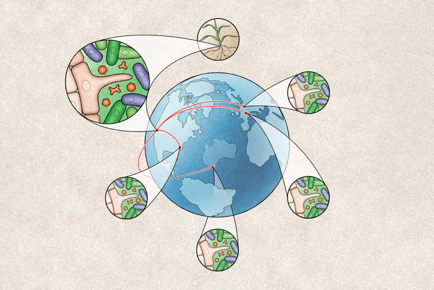
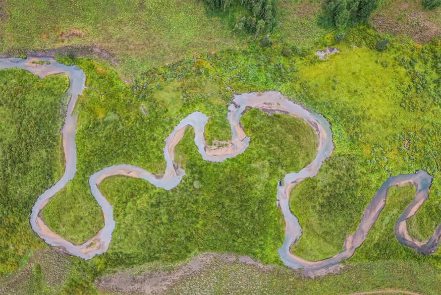
Cataloging the Microbiome of U.S. Rivers
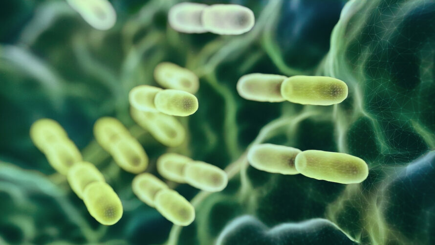
Revealing the Mysteries Within Microbial Genomes
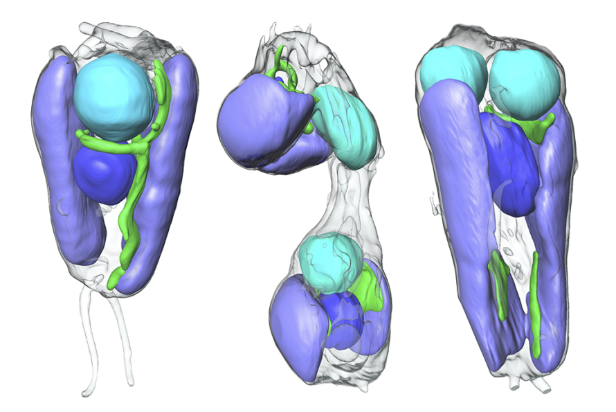
Scientists Discover First Nitrogen-Fixing Organelle
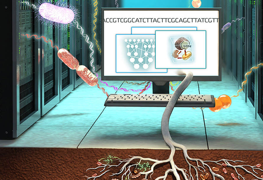
Improving Climate Predictions by Unlocking the Secrets of Soil Microbes
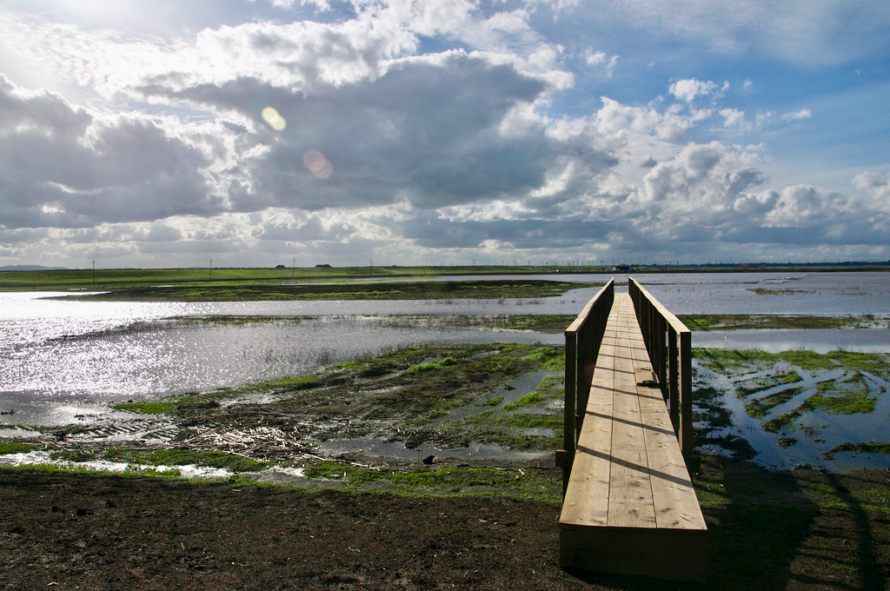
Rising Sea Levels Could Lead to More Methane Emitted from Wetlands
Basics 2 Breakthroughs
Doubling Down on Known Protein Families