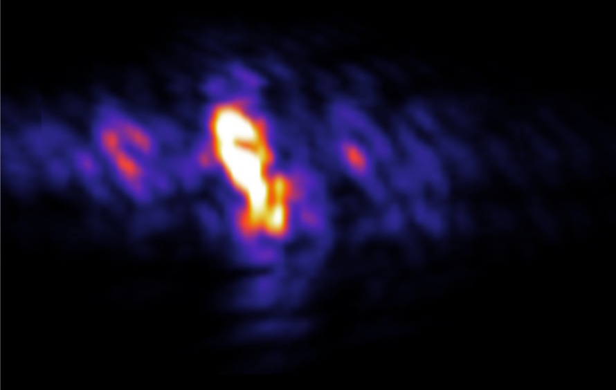
Atomic X-ray Laser Opens Door to Attosecond Imaging
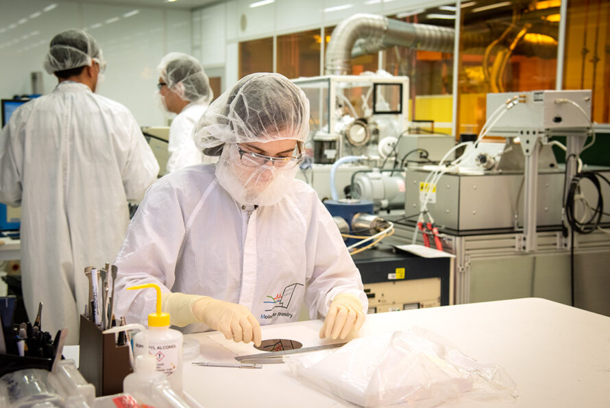
Six Scientific Advances Made Possible by Berkeley Lab’s Molecular Foundry
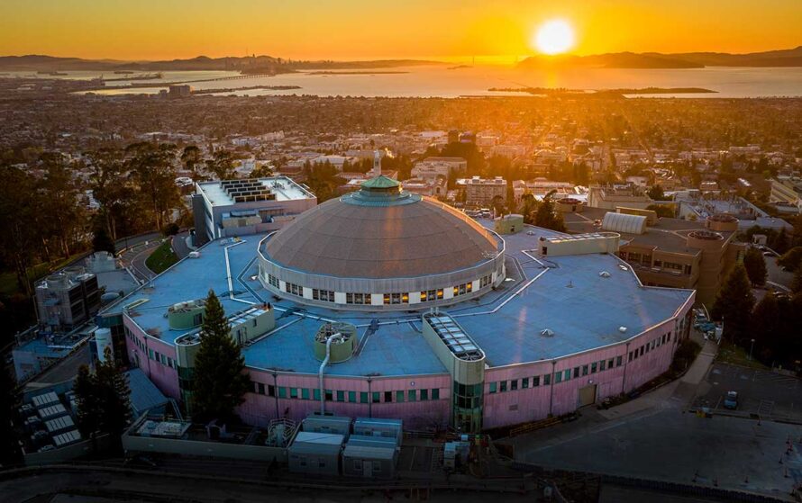
Bringing Discoveries to Light: Six Ways the Advanced Light Source Is Accelerating Technology Breakthroughs for Society
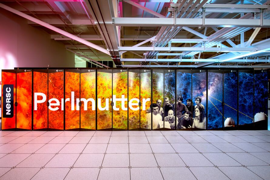
Five Ways Berkeley Lab's NERSC is Revolutionizing Scientific Research
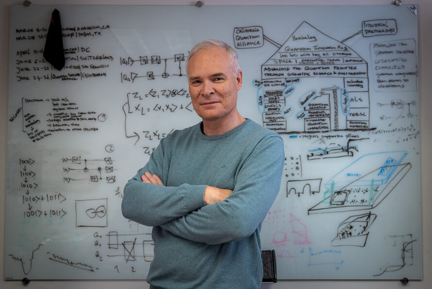
Science Power-up: Rewriting the Rules with Quantum Information Science
Seven Ways Berkeley Lab is Pioneering the Quantum Future
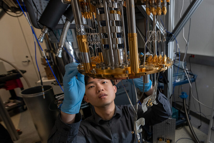
A Quiet Revolution: New Technique Could Accelerate Noise-Free Superconducting Qubits for Quantum Computing

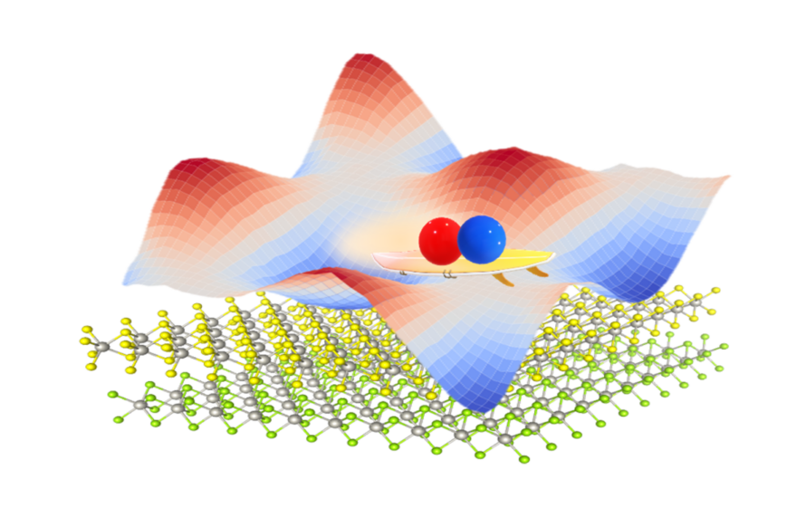
Moiré than Meets the Eye
For Better Quantum Sensing, Go With the Flow
U.S. Department of Energy National Quantum Information Science Research Centers Celebrate 4-year Milestone, Look Toward Future
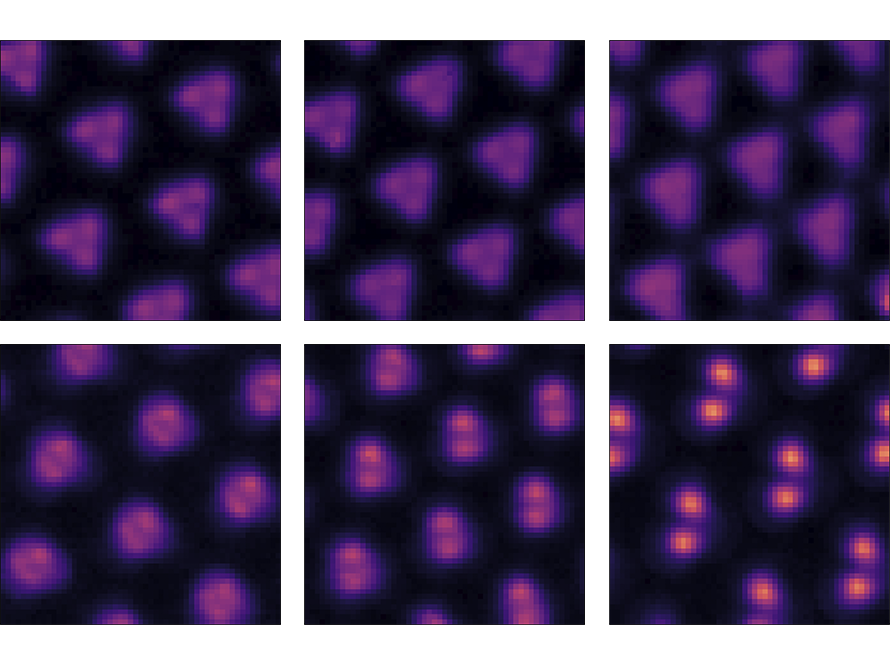
Scientists Capture Images of Electron Molecular Crystals
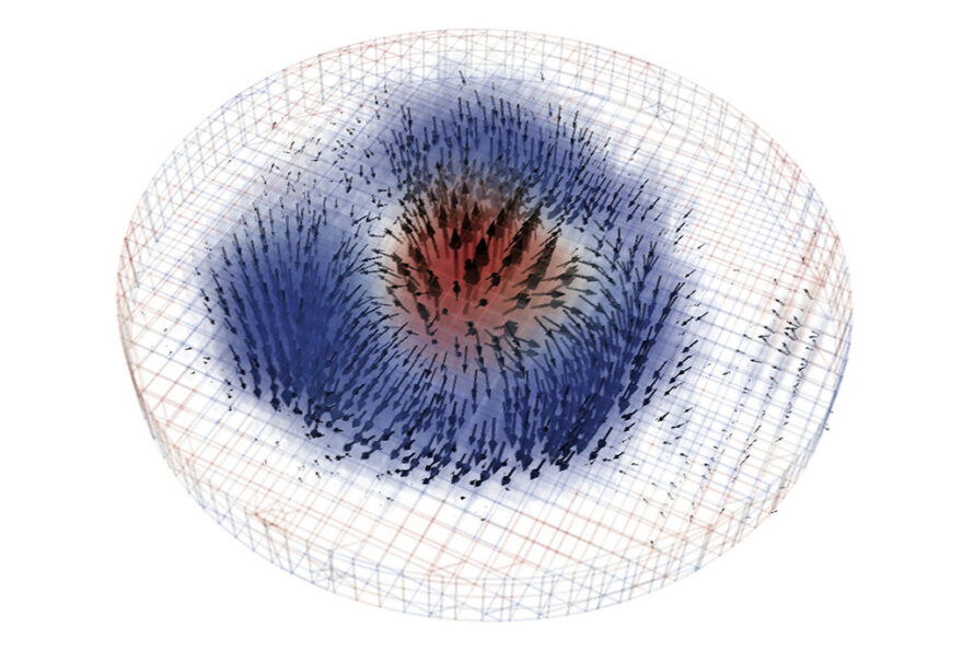
Researchers Succeed in Taking 3D X-ray Images of a Skyrmion