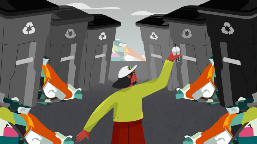
Putting an End to Plastic Separation Anxiety
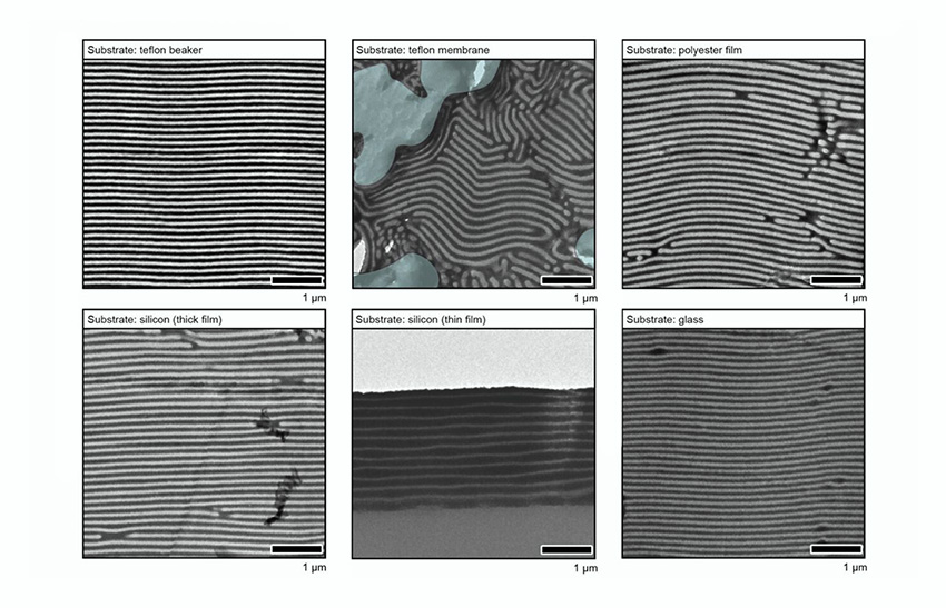
Scaling Up Nano for Sustainable Manufacturing
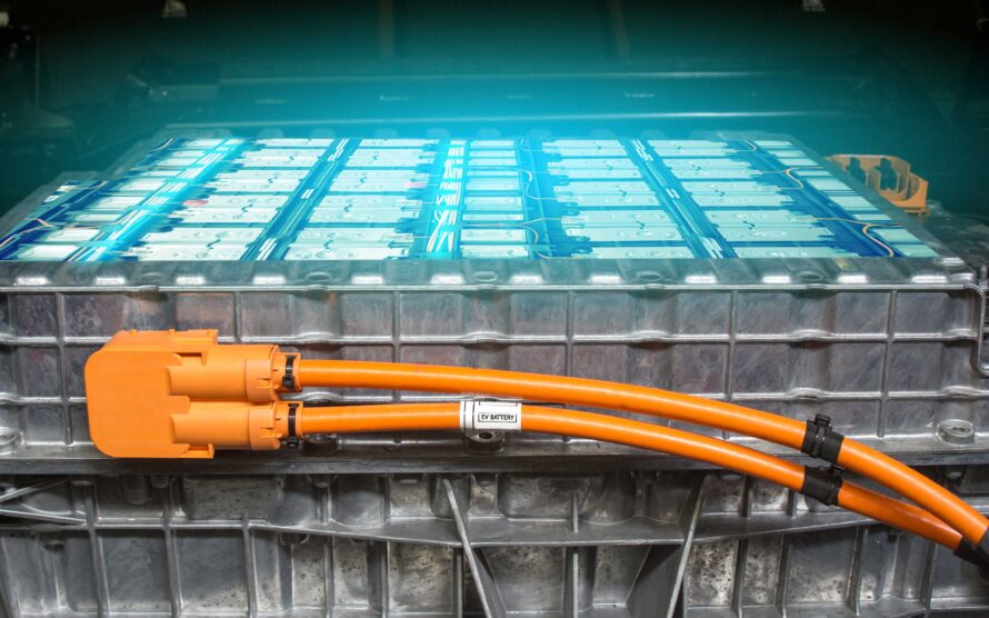
Why Do Batteries Sometimes Catch Fire and Explode?
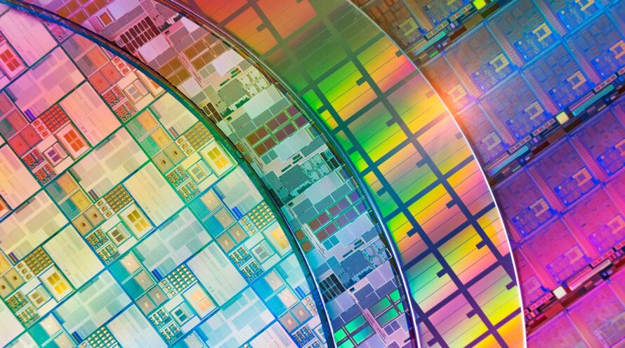
Accelerating Sustainable Semiconductors With ‘Multielement Ink’
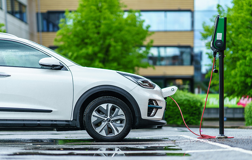
New Consortium to Make Batteries for Electric Vehicles More Sustainable
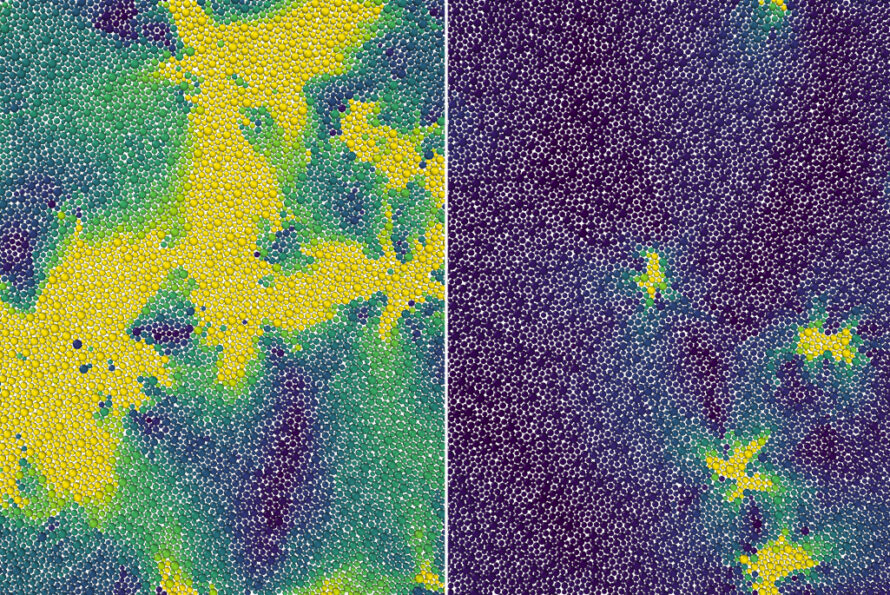
Scientists Theorize a Hidden Phase Transition Between Liquid and a Solid
Making Renewable, Infinitely Recyclable Plastics Using Bacteria
Watching Molecules Relax in Real Time
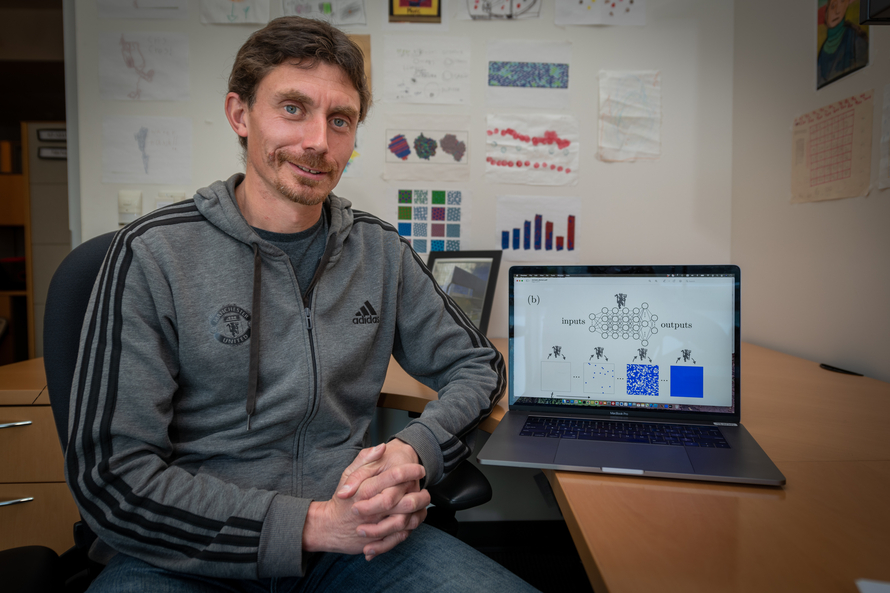
Harnessing Machine Learning to Make Complex Systems More Energy Efficient
Research on Light Emission From Black Phosphorus Hints at New Applications
Scientists Create a Longer-Lasting Exciton that May Open New Possibilities in Quantum Information Science
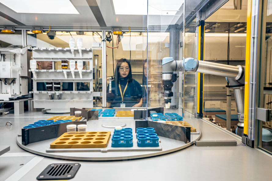
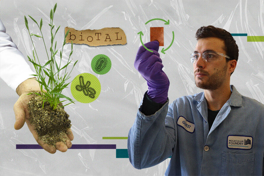
Meet the Autonomous Lab of the Future