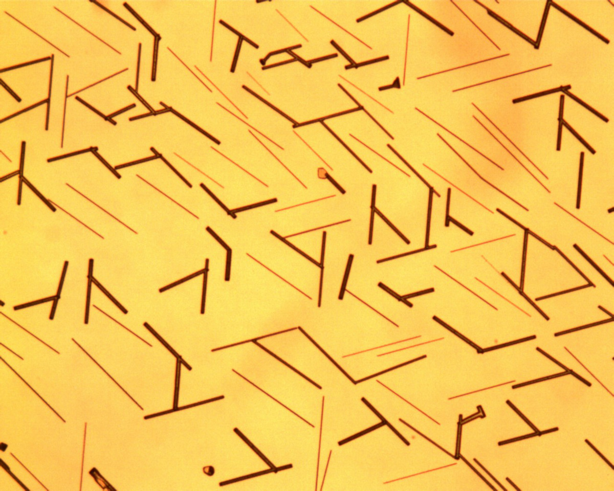
New Computer Simulations Help Scientists Advance Energy-Efficient Microelectronics

New Technology Provides Electrifying Insights into How Catalysts Work at the Atomic Level
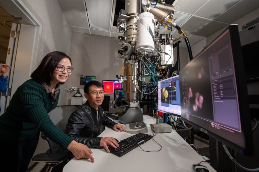
Groundbreaking Microcapacitors Could Power Chips of the Future
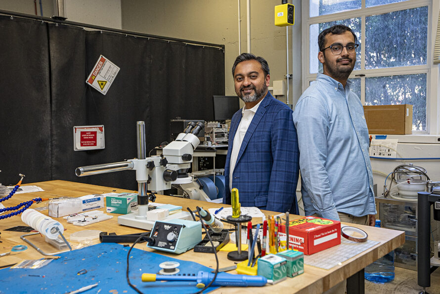
Two Berkeley Lab Researchers Elected to the National Academy of Sciences

New Technique Lets Scientists Create Resistance-Free Electron Channels

Scientists Advance Affordable, Sustainable Solution for Flat-Panel Displays and Wearable Tech
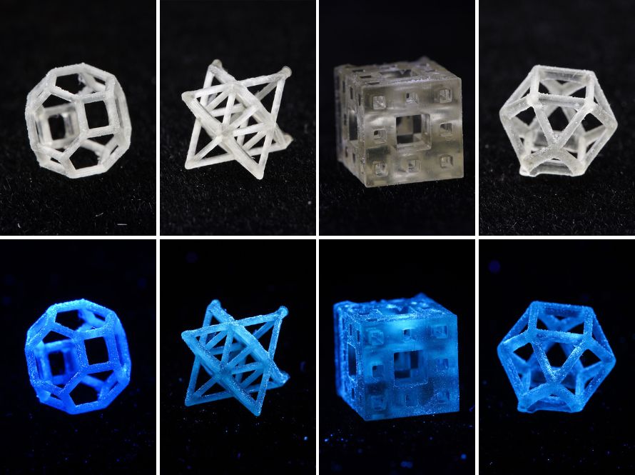
Scaling Up Nano for Sustainable Manufacturing
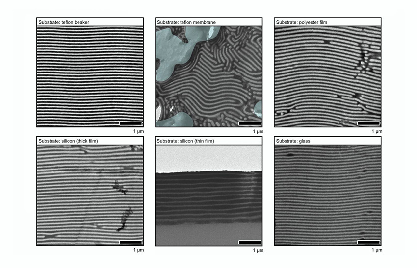
Accelerating Sustainable Semiconductors With ‘Multielement Ink’
How Scientists Are Accelerating Next-Gen Microelectronics
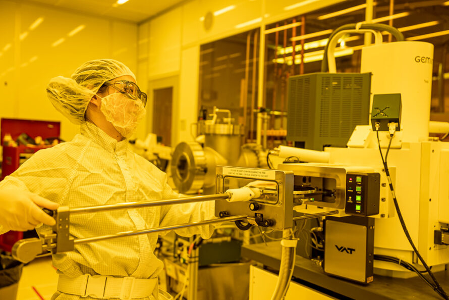
Electronic Bridge Allows Rapid Energy Sharing Between Semiconductors
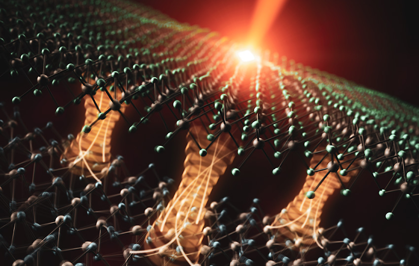
Science in Motion: Nano-Materials to Make Better Light Sensors

Scientists Grow Lead-Free Solar Material With a Built-In Switch
