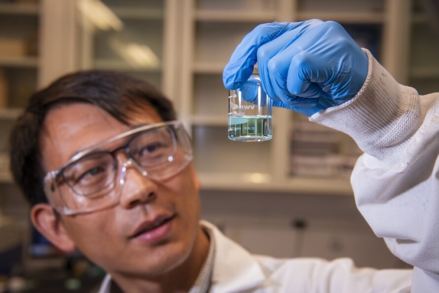
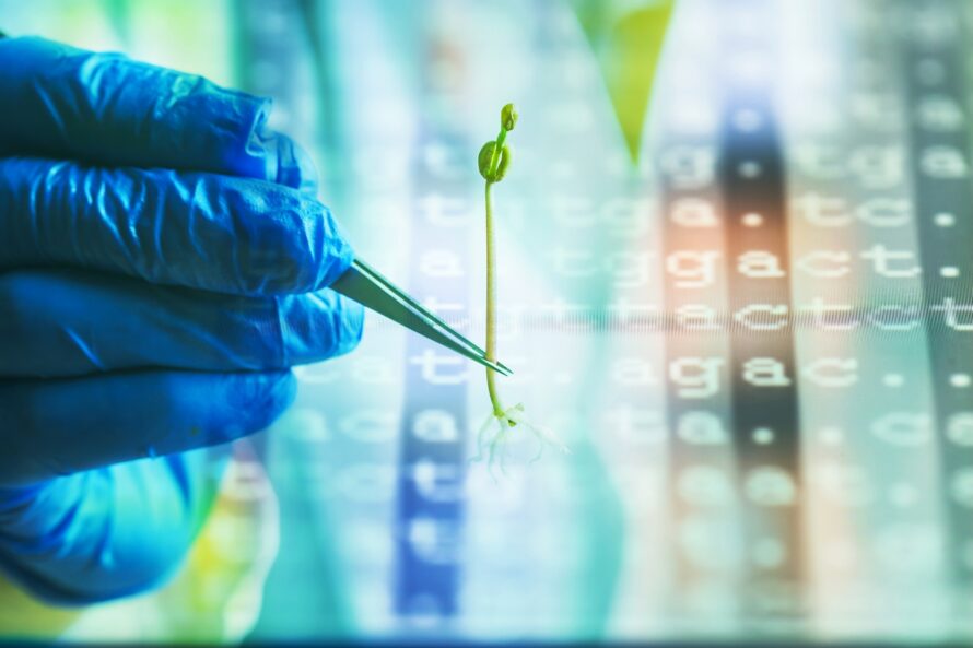
Scientists Build 'Speed Scanner' to Test Thousands of Plant Gene Switches at Once
Berkeley Lab’s Big Science Stories of 2025
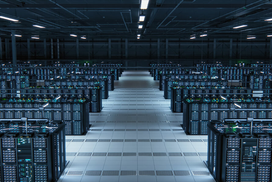
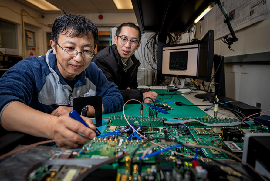
How Researchers Are Driving Advances for Data Centers
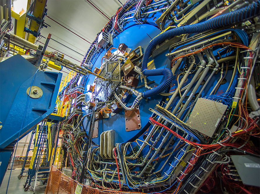
LZ Sets a World’s Best in the Hunt for Galactic Dark Matter and Gets a New Look at Neutrinos from the Sun’s Core
New Data Release from CUORE Features a “Noise-Canceling” Algorithm
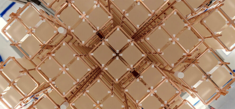
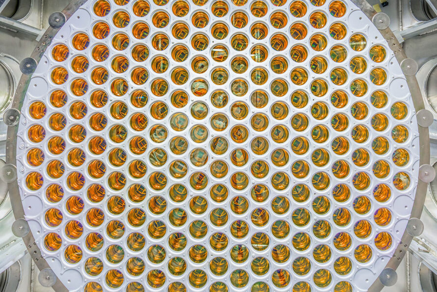
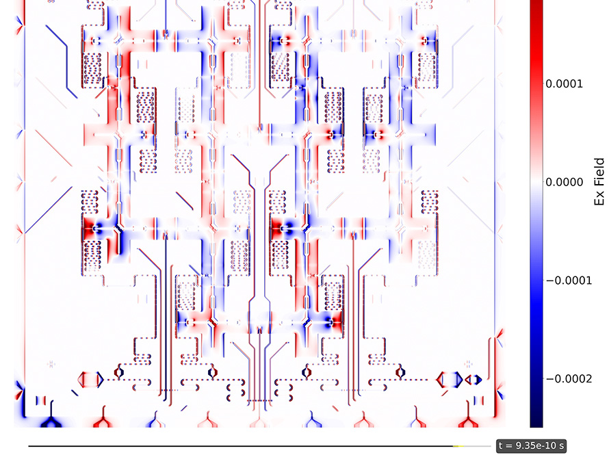
Unprecedented Perlmutter Simulation Details Quantum Chip
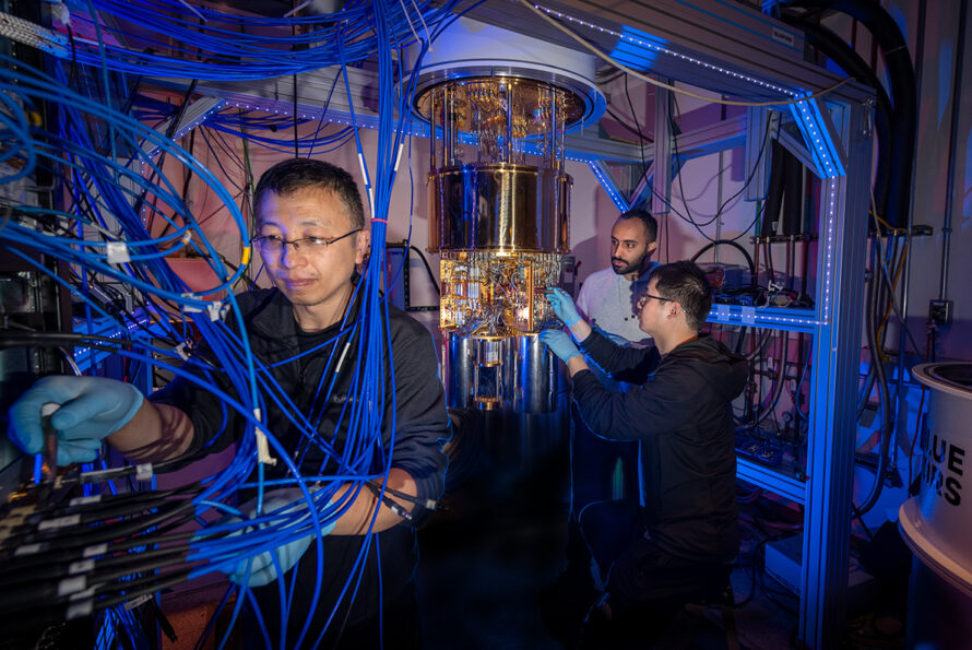
The Quantum Systems Accelerator Embarks on Next Five Years of Pioneering Quantum Technologies for Science
New Berkeley Lab and NVIDIA Partnership Integrates Quantum and AI Supercomputing for Next-Generation Research
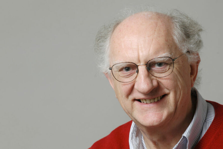
Former Berkeley Lab Scientist John Clarke Wins 2025 Nobel Prize in Physics
More Signs of Phase-change ‘Turbulence’ in Nuclear Matter
Atomic Neighborhoods in Semiconductors Provide New Avenue for Designing Microelectronics
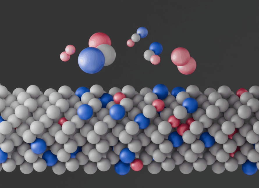
Electron Microscopy Reveals New Method to Make Exotic Metal Alloys