India Can Achieve Energy Independence by 2047 Through Clean Technology: Study

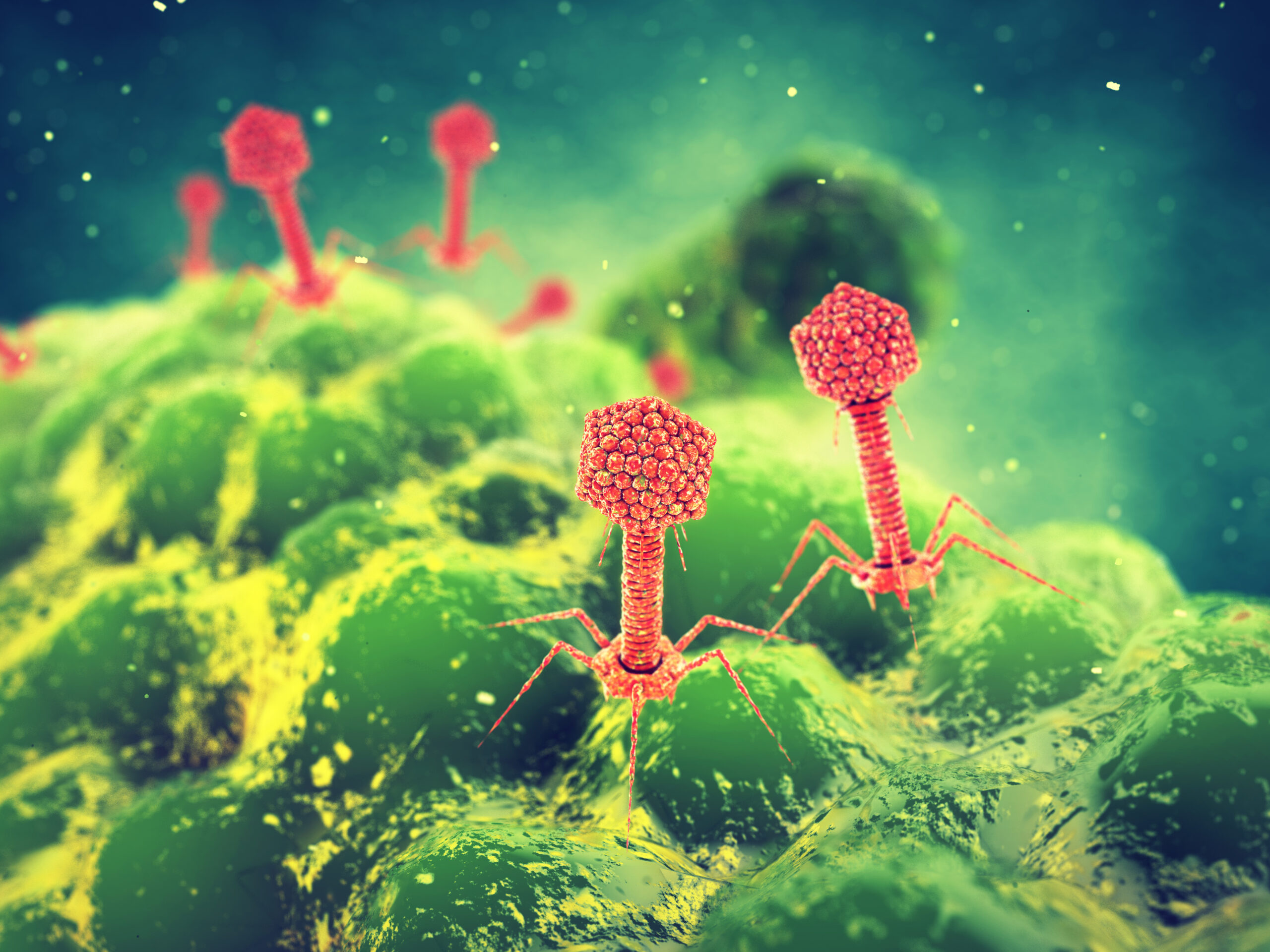
A Quick New Way to Screen Virus Proteins for Antibiotic Properties
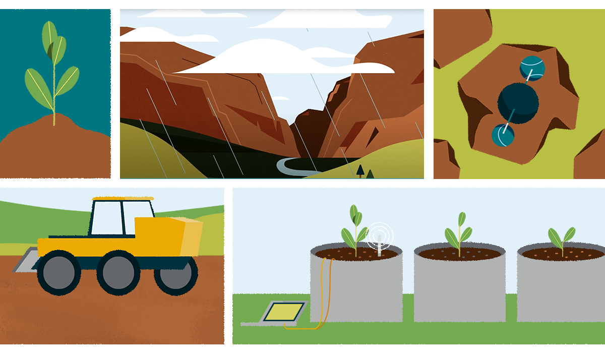
Science in Motion: Can enhanced weathering help slow climate change?
A Day in the Half-Life

Electric Vehicle Batteries Could Get Big Boost With New Polymer Coating

New DESI Planetarium Show to Premiere in Spring 2023

Berkeley Lab Director Mike Witherell to Serve on Governing Council of the National Academy of Sciences

On the Road to Better Solid-State Batteries
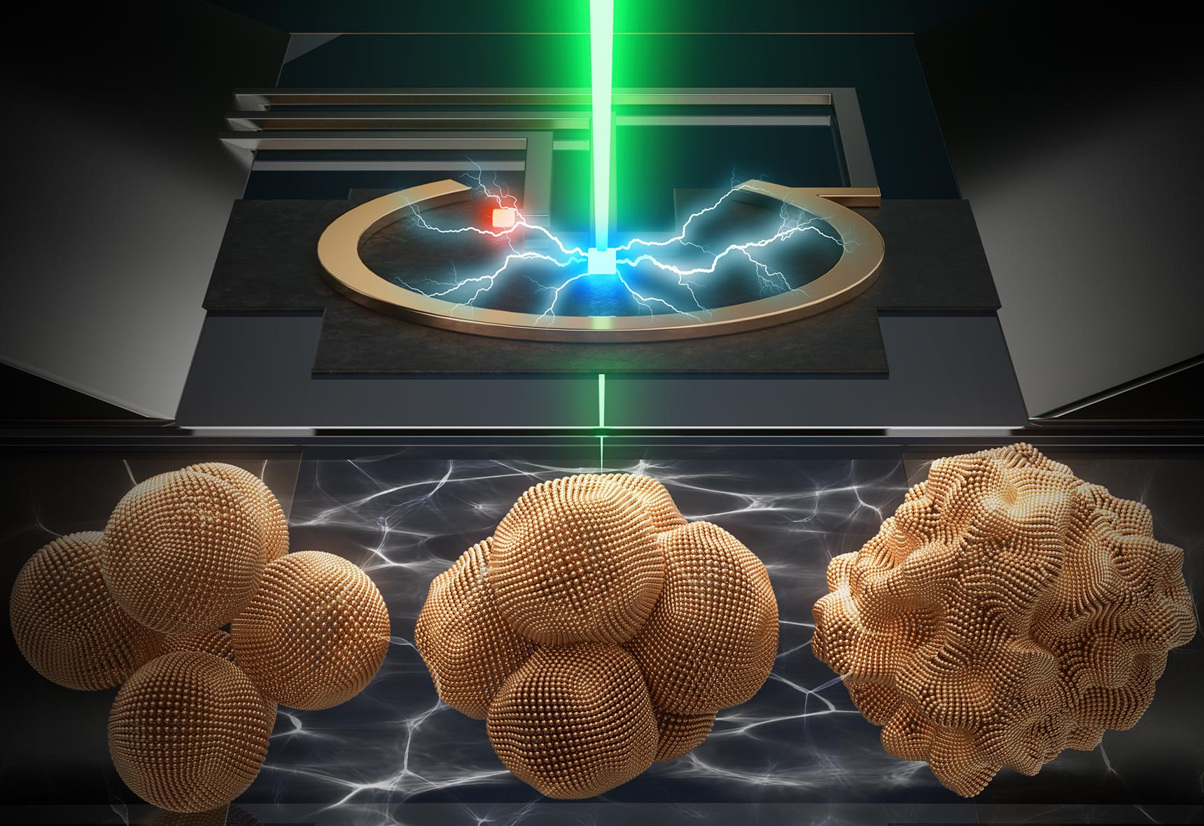
How a Record-Breaking Copper Catalyst Converts CO2 Into Liquid Fuels
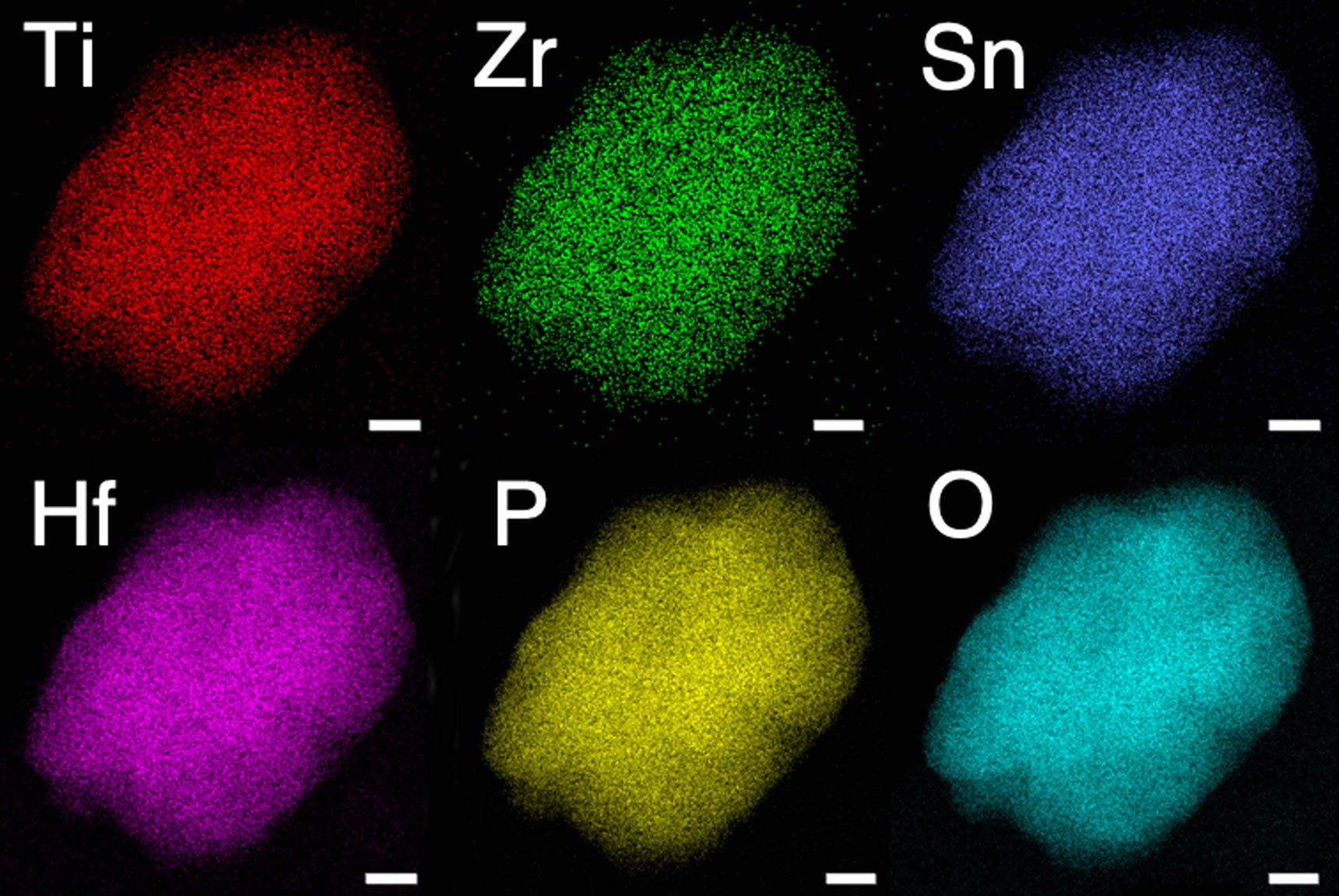
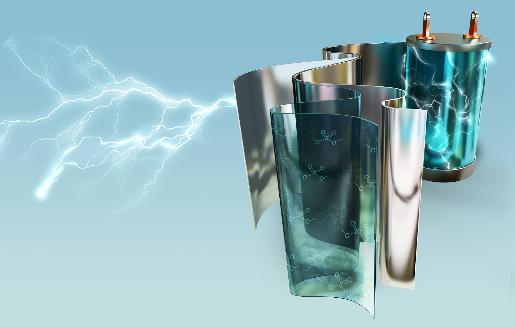
New Compound That Withstands Extreme Heat and Electricity Could Lead to Next-Generation Energy Storage Devices
Doubling Protected Lands for Biodiversity Could Require Tradeoffs With Other Land Uses, Study Finds

The Most Advanced Bay Area Earthquake Simulations Will be Publicly Available